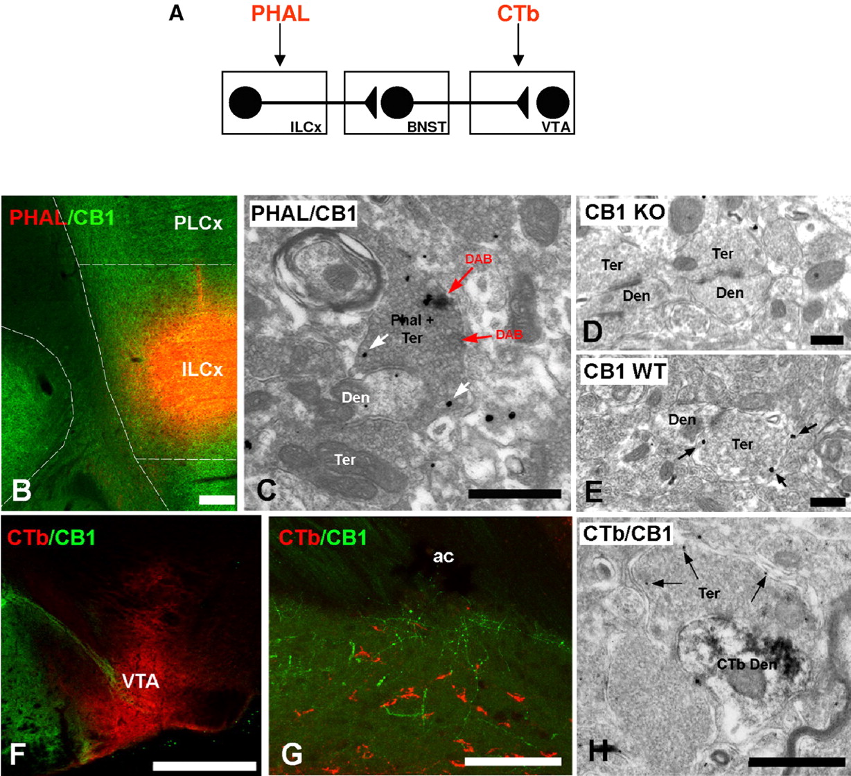
Immunolocalization of CB1-R in the BNST after injection of tracers in the prefrontal cortex (PHA-L) and mesencephalon (CTb; scheme in A). B, Double immunofluorescence confocal for the injection site of the tracer PHA-L (red) injected in the ILCx (scheme on top of the figure) and the expression of CB1-R (green). Scale bar, 500 μm. C, Electron micrograph showing colocalization of transported PHA-L and immunoreactivity for CB1-R in terminals in BNST using preembedding immunoperoxidase and immunogold methods, respectively. Cortical axons identified by cytoplasmic DAB deposits (PHA-L plus Ter, red arrow) have CB1-R immunoparticles on their membranes (white arrows). Scale bar, 0.3 μm. D, E, CB1-R deficient mice (CB1 KO) were used for control of antibody specificity. CB1-R immunolabeling was not observed in the BNST of CB1 KO. In wild-type mice (CB1 WT), CB1-R silver-intensified gold particles were localized along membranes of presynaptic bouton. Scale bar, 0.4 μm. F, Injection site of the retrograde tracer CTb was restricted to the VTA. Scale bars, 100 μm. G, Double immunofluorescence confocal for the tracer CTb (red) injected in the VTA and the expression of CB1-R (green) in BNST. Scale bar, 100 μm. H, Subcellular distribution of CTb and CB1-R in BNST using preembedding immunoperoxidase and immunogold methods, respectively, for electron microscopy. Immunodeposits of the retrogradely transport CTb were on dendrites (Den) of BNST projecting neurons to VTA (CTb plus Den) apposed to a CB1-R immunopositive synaptic terminal (Ter) (arrows, CB1-R gold particles) and making an asymmetrical synapse with a CB1-R immunonegative Ter. Scale bar, 0.3 μm. ac, Anterior commissure.


