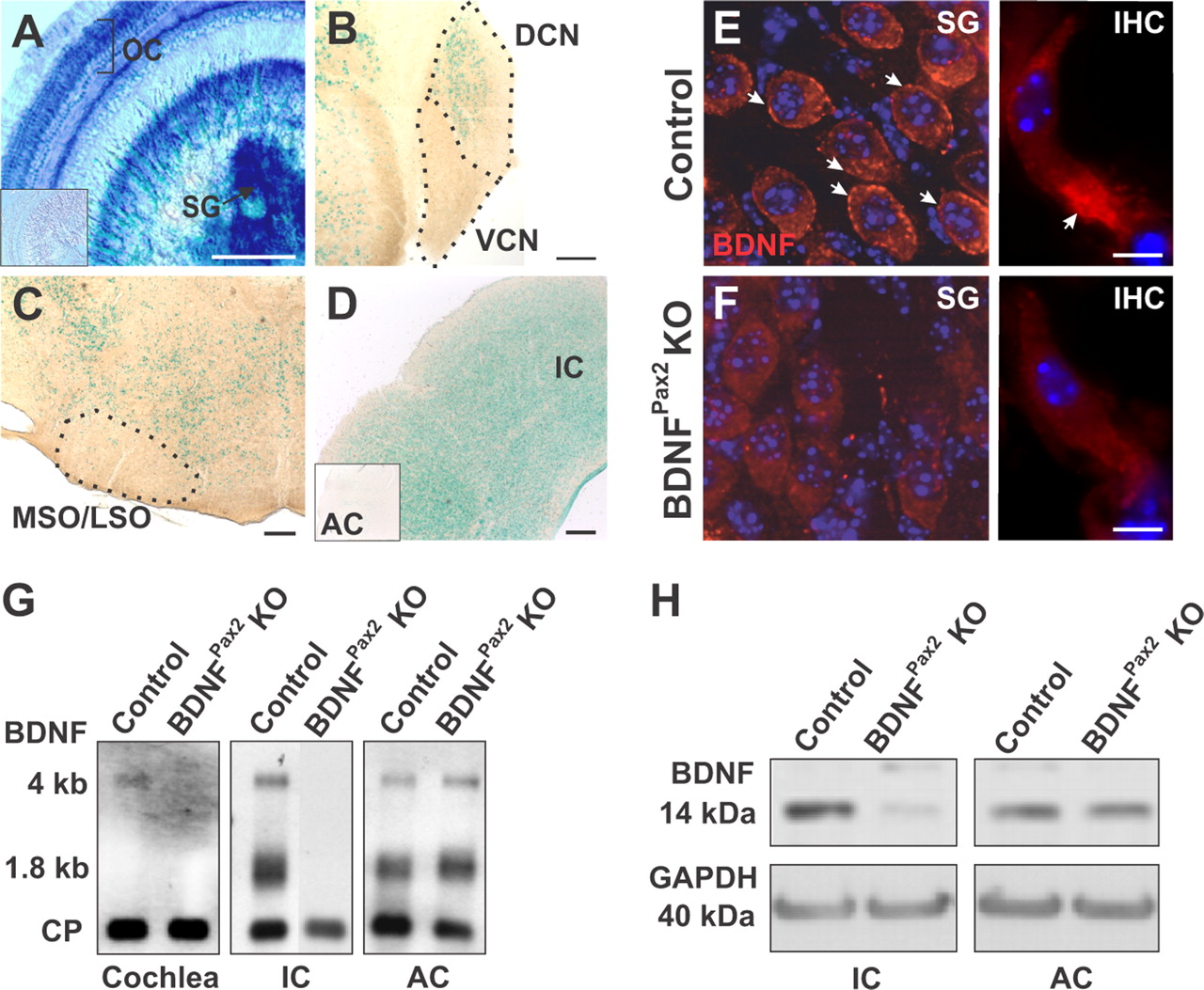
Inactivation of BDNF in the auditory system. A–D, X-Gal staining of mice carrying the Pax2-Cre transgene on a ROSA26R background (Soriano, 1999), revealed Cre activity in the mature cochlea (A, organ of Corti, OC, and spiral ganglion neurons, SG), the dorsal (but not ventral) cochlear nucleus (B, DCN, VCN) and inferior colliculus (D, IC). No staining was observed in the olivary complex (C, MSO/LSO) and in all layers of the auditory cortex (D, inset, AC). Scale bar, 200 μm; n = 4. E, F, Immunohistochemistry for BDNF (red) on cochlear sections from control (E), BDNFPax2 KO (F) mice at the level of SG (left) and IHCs (right). Cell nuclei were counterstained with DAPI, n = 3 mice, done in triplicate. Scale bar, 10 μm. G, Northern blot of mRNA from cochlea, IC and AC tissue of control and BDNFPax2 KO mice, hybridized with a probe for BDNF exon IX recognizing BDNF mRNA isoforms (1.8 kb and 4 kb; Timmusk et al., 1993). Cyclophilin (CP) was used as reference gene, n = 4 mice, done in duplicate. H, Western blot detection of BDNF in IC and AC from control and BDNFPax2 mice. GAPDH was used as loading control, n = 4 mice, done in duplicate.


