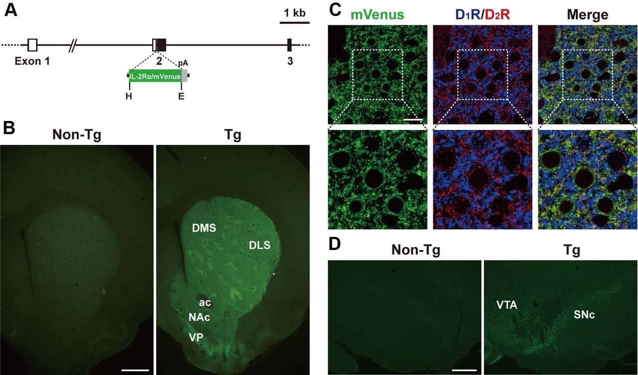
Generation of Tg rats expressing IT receptor under the control of the Drd2 gene promoter. A, Structure of the transgene construct. The gene cassette encoding IL-2Rα/mVenus followed by the SV40 early gene polyadenylation signal (pA) was introduced into the place of the initiation codon in exon 2 of the rat Drd2 gene. Exons 1–3 of the rat Drd2 gene are shown. Open and closed boxes indicate the 5′-noncoding and coding regions, respectively. E, EcoRI; H, HindIII. B, Detection of endogenous fluorescence in sections through the striatum prepared from Tg and non-Tg rats. Transgene expression is observed in the DLS, DMS, NAc, and ventral pallidum (VP) in the Tg rats. ac, anterior commissure. C, Triple immunofluorescence histochemistry for mVenus, D1R, and D2R with sections through the DLS. mVenus-, D1R-, and D2R-positive signals are shown in green, blue, and red, respectively. In a merged image, green signals overlap with red signals, and thus emit yellow signals. Lower images are magnified views of squares in upper images. D, Immunofluorescence histochemistry for mVenus with sections through the ventral midbrain. Transgene expression is observed in the SNc and VTA in the Tg rats. Scale bars: B, 1 mm; C, 20 μm; D, 400 μm.


