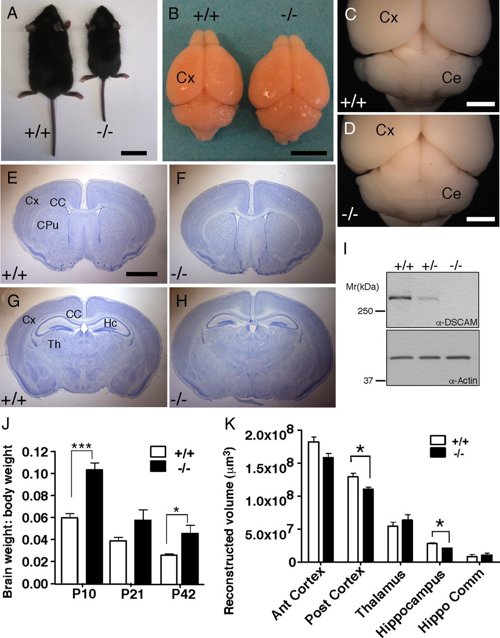
DSCAMdel17 mutants exhibit abnormal gross brain morphology. A–I, At postnatal day P21, DSCAMdel17 mutant mice are smaller in body size compared with their wt littermates (A). The brains of DSCAMdel17 mutants are increased in length and decreased in width (B), have an enlarged tectum and abnormally shaped cerebellum (B, D) compared with wt littermates, B and C. E–H, Nissl-stained coronal sections (50 μm) of corresponding regions in wt at different levels, E and G, and mutant brains, F and H. In DSCAM del17 mice the total brain size is increased whereas the hippocampal (HC) size is decreased (H), compared with wt (G). I, Protein expression in tissue extracts derived from the cortex of wt, DSCAMdel17 heterozygote, or mutant mice. DSCAM protein expression accessed by Western blotting in cortical extracts derived from P21 brains of the indicated genotype. α-Actin was used as a loading control. J, Ratio of brain to body weight at P10, P21, and P42 in wt and DSCAMdel17 mutant mice. Analysis was performed on the following: P10: wt, n = 5; DSCAMdel17, n = 3; P21: wt, n = 6; DSCAMdel17, n = 3; P42: wt, n = 5; DSCAMdel17, n = 3. K, Volume reconstruction of defined areas of P21 anterior cortex (Ant cortex), posterior cortex (Post cortex), thalamus (Th), hippocampus (Hippo), and the hippocampal commissure (Hippo Comm). Brains for each genotype were evaluated: n = 3. CPu, Caudate–putamen; Th, thalamus. Scale bars: 2 cm (A), 500 mm (B), 2 mm (C–H). Mean ± SE; *p < 0.05, ***p < 0.001 compared with wt (Student's t test).


