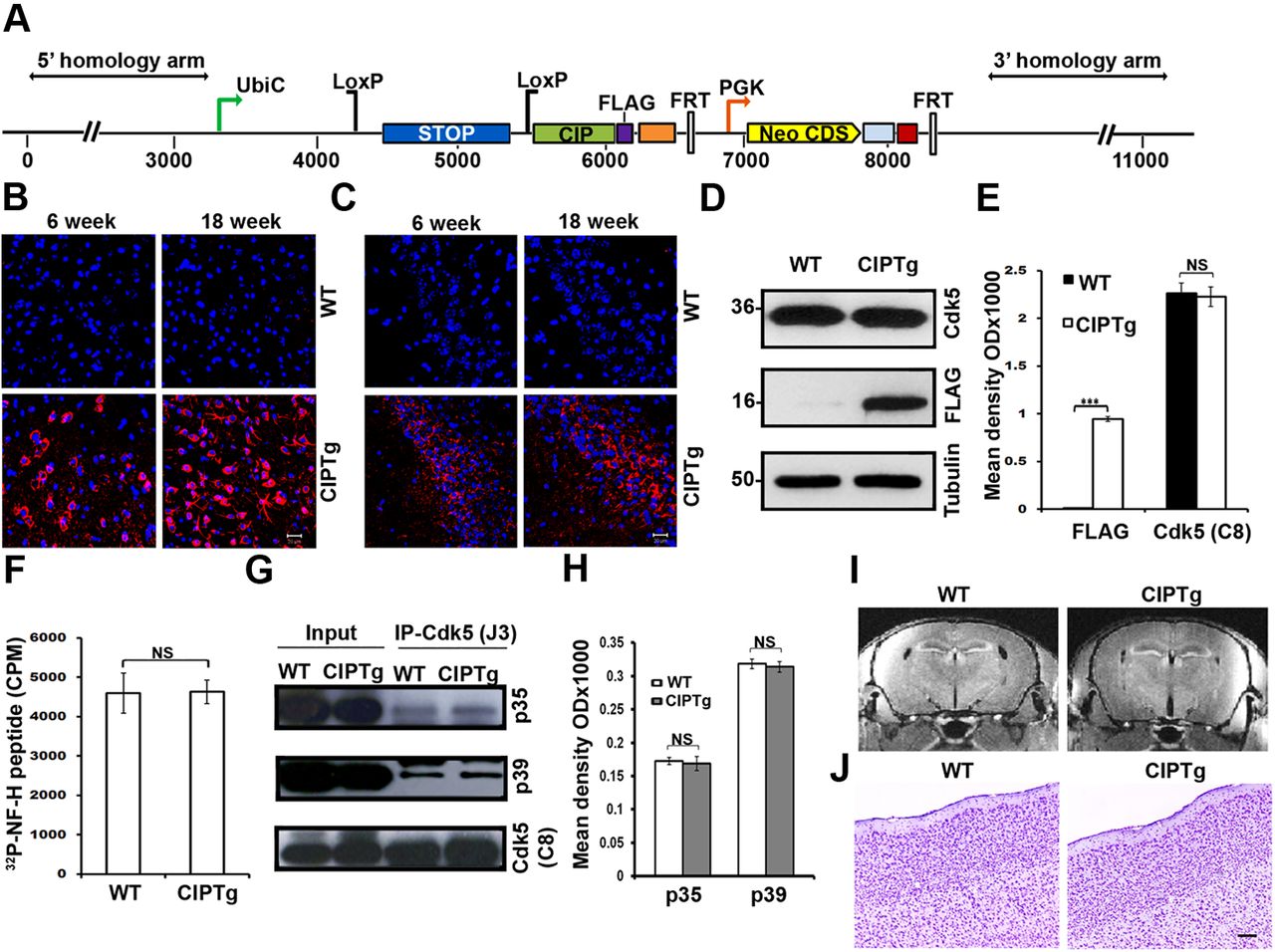
Characterization of CIPTg mice. A, The CIPTg construct contains a 3′-FLAG-tagged CIP transgene incorporated in to the ROSA26 locus whose 5′ regulatory elements were separated from the coding region with a floxed stop sequence. A frt-flanked neomycin resistance (Neo) cassette was inserted next to the transgene before the 3′ homology arm. Representative confocal images of frontal cortex (layer 2/3) (B) and hippocampus (CA3 region) (C) from 6-week-old and 18-week-old WT and CIPTg mice. The sections were immunostained with anti-FLAG antibody (red), and nuclei were counterstained with DAPI (blue). D, Immunoblot analyses of brain lysates from WT and CIPTg mice using anti-Cdk5 (C8) and anti-FLAG antibodies. Equal amounts of protein loading were confirmed by reprobing the membrane with anti-tubulin antibody. E, Quantification of immunoblot analyses in D by densitometric scanning (n = 3) (***p < 0.001 and NSp > 0.05). F, Kinase assays using active kinase (Cdk5) from WT and CIPTg mice brains to phosphorylate a high-molecular-weight neurofilament (NF-H) peptide (NSp > 0.05). G, WT and CIPTg mice brain lysates were immunoprecipitated (IP) using anti-Cdk5 (J3) antibody. Samples were separated by SDS-PAGE and immunoprobed with anti-p35, anti-p39, and anti-Cdk5 (C8) antibodies. H, Quantification of immunoblot analyses in G by densitometric scanning (NSp > 0.05). MRI scanning images of coronal slices (I) and Nissl staining images of frontal cortex (J) from WT and CIPTg mice brains. Scale bars, 20 μm. Data are representative of n = 4 mice. Error bars indicate ±SEM.


