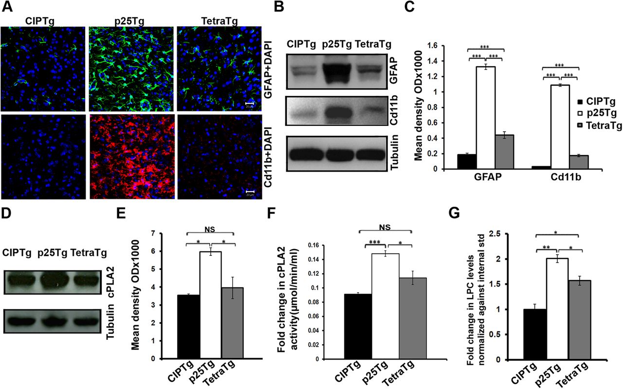
Reduced neuroinflammation in TetraTg mice. A, Representative confocal images of frontal cortex from 12 week induced p25Tg, TetraTg, and CIPTg mice. The brain sections were labeled with anti-GFAP antibody (green), anti-Cd11b antibody (red), and nuclei were stained with DAPI (blue). B, Western blot analyses of brain lysates from p25Tg, TetraTg, and the CIPTg mice using anti-GFAP and anti-Cd11b antibodies. C, Quantification of immunoblots in B by densitometric scanning (***p < 0.001). D, Immunoblot analyses were performed on lysates from the samples same as in B using anti-cPLA2 antibody. E, Quantification of immunoblot analyses in D (*p < 0.05, NSp > 0.05). F, cPLA2 activity assays were performed with lysates from p25Tg, TetraTg, and CIPTg mice (*p < 0.05, NSp > 0.05, ***p < 0.001). G, Results from the mass spectrometric analyses for the p25Tg, TetraTg, and CIPTg mice brain samples. Results were normalized against the internal standards of LPC (*p < 0.05, **p < 0.01). Scale bars, 20 μm. Data are representative of n = 4 mice. Error bars indicate ±SEM.


