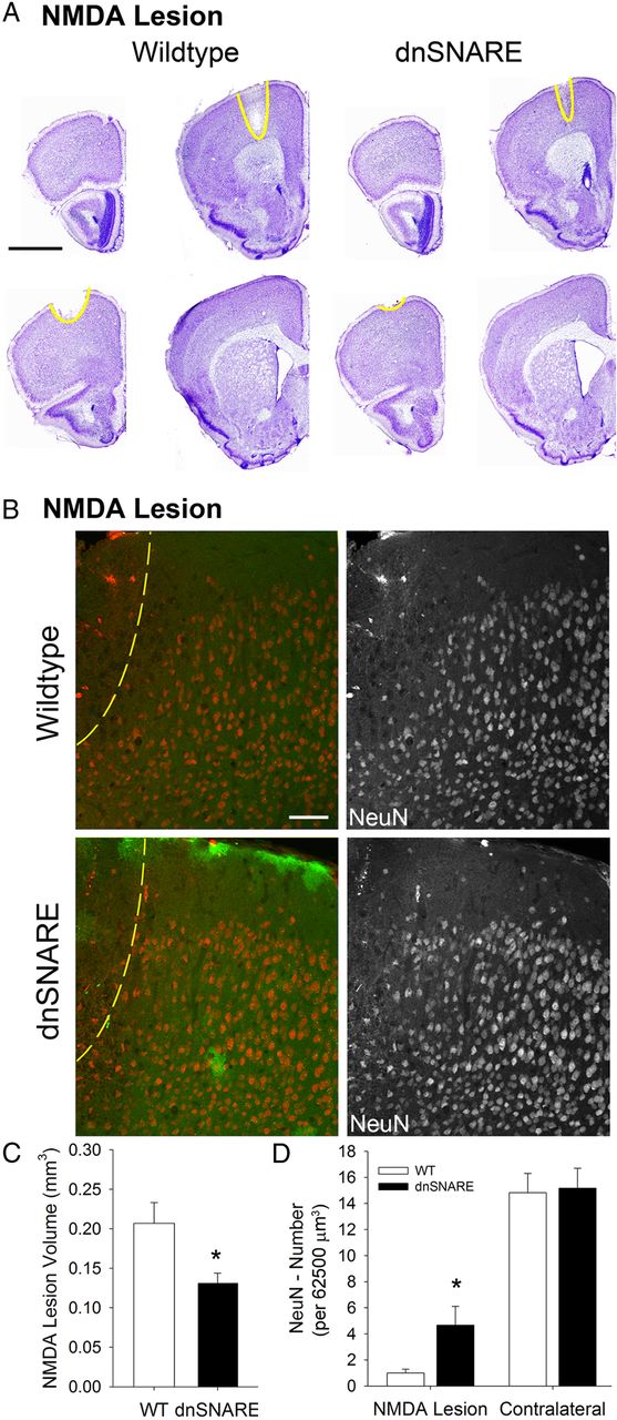
Figure 4.
Damage following NMDA lesions is attenuated by dnSNARE expression in astrocytes. A, Representative images of Nissl-stained sections from wild-type and dnSNARE mice subjected to unilateral NMDA lesions. B, Representative image of NeuN (red) staining in wild-type and dnSNARE mice subjected to unilateral NMDA lesions. C, Quantification of NMDA lesion volume calculated from reconstructed Nissl-stained serial section sets in wild-type and dnSNARE mice. D, Quantification of the number of NeuN-positive neurons per 62,500 μm3 in wild-type and dnSNARE mice in NMDA lesion and contralateral cortical tissue. *p < 0.05.


