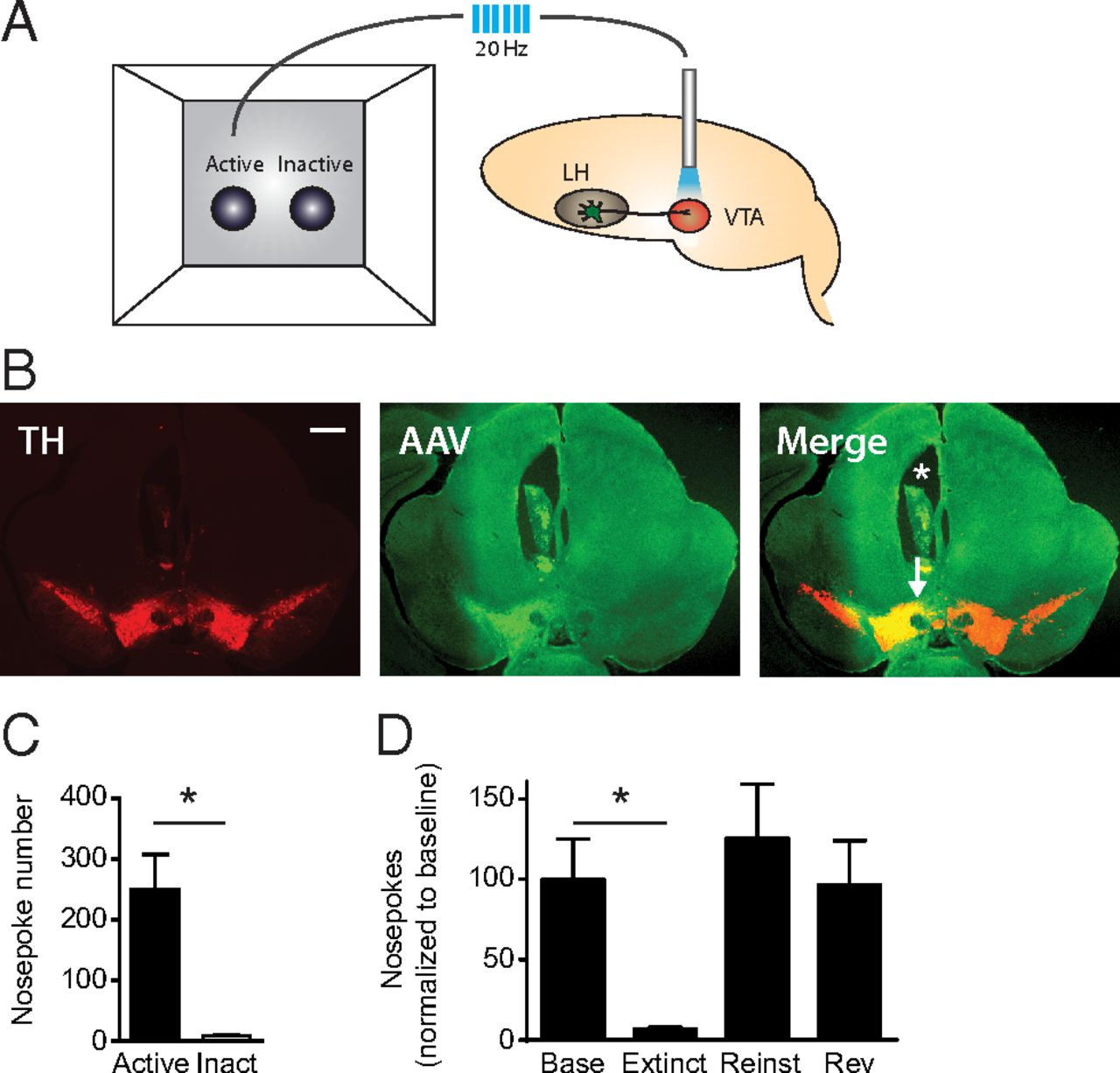
A, Schematic of the optical ICSS paradigm. Each active nosepoke produced a train of 20 Hz blue light pulses (5 ms each) to stimulate lateral hypothalamic axons terminating in the VTA. B, Images showing cannula placement aimed at the VTA. TH (red) indicates midbrain dopamine neurons; green represents ChR2-expressing neurons (AAV). In the merged image: *The cannula location. The arrow indicates cannula path toward the VTA. Scale bar, 500 μm. C, Average nosepoke rates for optical stimulation of LH to VTA fibers. Mice perform significantly more active nosepokes (black) than inactive nosepokes (white) (p < 0.001, n = 10, Mann–Whitney test). D, Extinction experiment depicting operant responding at the active nosepoke. Base, Baseline session to which active nosepokes were normalized; Extinct, extinction experiments in which the laser was turned off 10 min into the hour-long trial, nosepoke cues remained present; Reinst, reinstatement sessions in which each active nosepoke produced optical stimulation in the absence of nosepoke cues; Rev, reversal experiments during which the active and inactive nosepokes were switched. Active nosepokes during extinction, but not reinstatement or reversal, were significantly lower than baseline (p = 0.006, n = 10, Dunnett's multiple-comparisons test).


