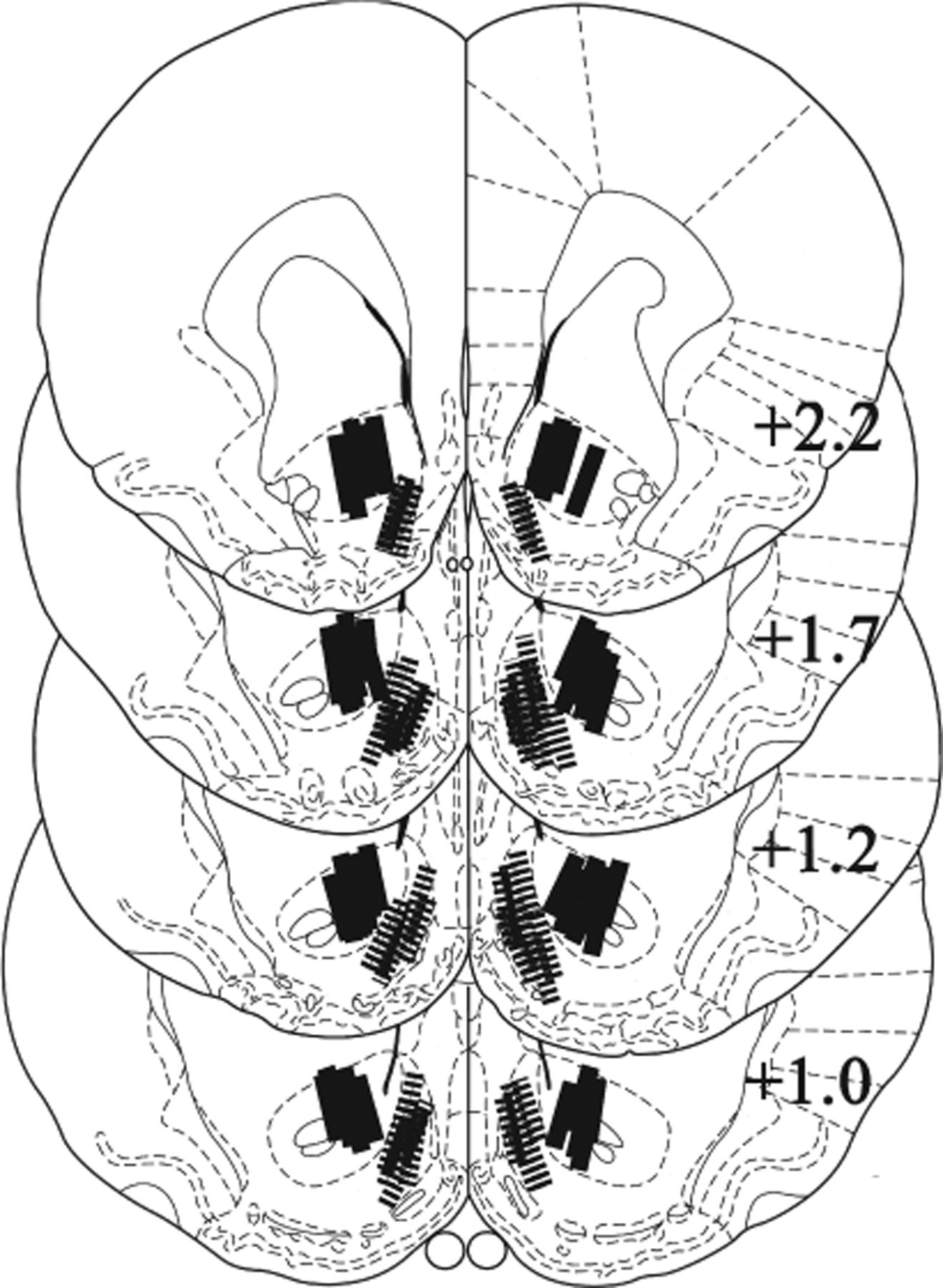
Figure 2.
Localization of probe placements. Only the location of the active dialysis membrane (1.0 mm) is shown. Note that the probes in the shell region (hatched bars) are angled from the opposite hemisphere; this procedure avoids penetration of the ventricle and involves about the same tissue damage (Suto et al., 2009, 2010; Suto and Wise, 2011) as is seen with the core probes (solid bars), which are angled from a more lateral entry. The numbers indicate the distance (millimeters) posterior to the bregma (Paxinos and Watson, 2007).


