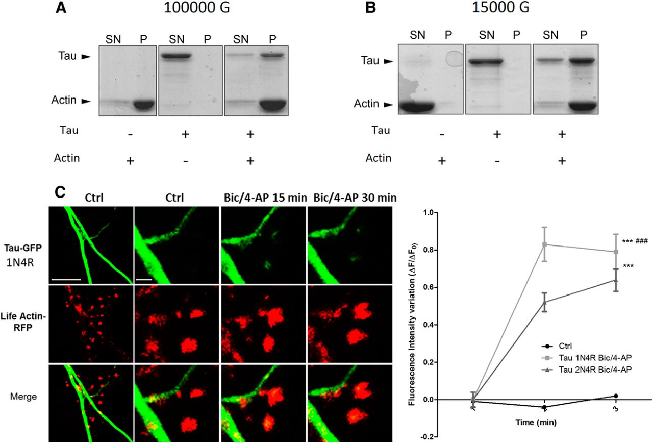
Tau interacts with actin in purified system. Cosedimentation of actin filaments with tau. Actin was polymerized at 5 μm for 1 h at room temperature in the absence or in the presence of 1 μm tau. The reaction mixtures were then centrifuged at 100,000 × g to sediment actin filaments (A) or at 15,000 × g to precipitate only actin bundles (B). Supernatants (SN) and pellets (P) were applied to 10% SDS-PAGE gel and proteins were stained with Coomassie blue. C, Confocal imaging of cortical neurons (14 DIV) cotransfected with EGFP-Tau1N4R and LifeAct-RFP. Left, Spatial repartition of EGFP-Tau1N4R (green) and LifeAct-RFP (red) in untreated neurons. Scale bar, 5 μm. Right, Higher magnification of EGFP-Tau1N4R and LifeAct-RFP in a dendritic spine neuron after Bic/4-AP treatment (15 and 30 min, designated box in the left) by 15 and 30 min of Bic/4-AP treatment. The merged image shows colocalization of F-actin and tau. Scale bar, 1 μm. D, Quantification of fluorescence intensity mediated by EGFP-Tau1N4R and Tau2N4R in the head of spines during synaptic activation (Bic/4-AP treatment). The graph represents the evolution of ΔF/F0 generated by EGFP-Tau2N4R and EGFP-Tau1N4R in neurons subjected to synaptic activation or not. Statistical analysis was performed by two-way ANOVA followed by Bonferroni's posttest (mean ± SEM, ***p < 0.0001 vs control, ###p < 0.0001 vs Tau-2N4R; control n = 40 spines on 3 independent cultures; Bic/4-AP Tau2N4R n = 162 spines on 5 independent cultures, Bic/4AP Tau 1N4R n = 54 spines on 2 independent cultures).


