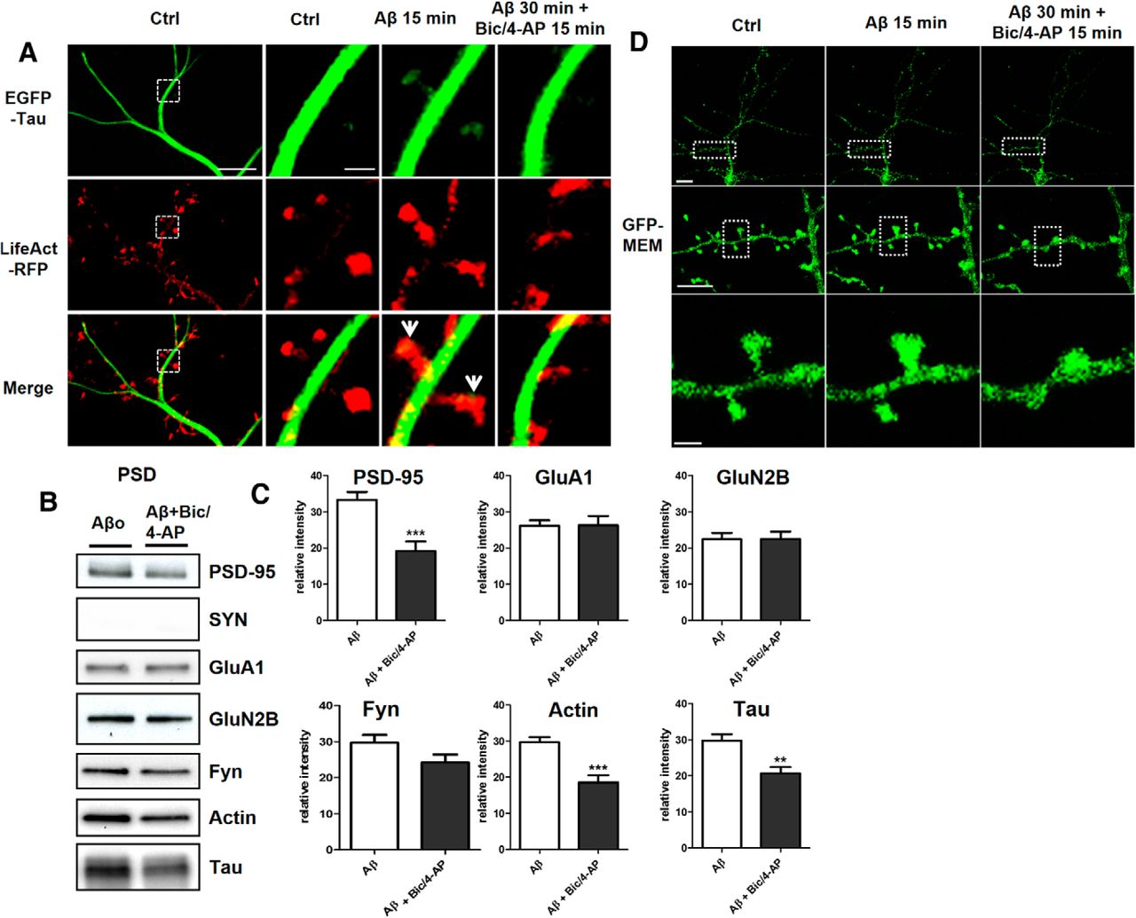
Aβo treatment on primary cortical neuron cultures induces synaptic mislocalization of endogenous tau that is disrupted by synaptic activation. A, Confocal imaging of cortical neurons (14 DIV) cotransfected with EGFP-Tau and LifeAct-RFP. Left, Spatial repartition of EGFP-Tau (in green) and LifeAct-RFP (in red) in untreated neurons. Scale bar, 5 μm. Right, Higher magnification of EGFP-Tau and LifeAct-RFP in a dendritic spine (designated box in the left) neuron showing the effect of 15 min of Aβ on tau localization (green) and actin cytoskeleton (red) followed by 15 min of Bic/4-AP. Scale bar, 1 μm. B, Representative Western blots of protein expression from fractionated primary cortical neurons treated with Aβo (100 nm) and then for 15 min with Bic/4-AP. Presynaptic protein (synaptophysin), postsynaptic proteins (PSD-95, GluA1,GluN2A, GluN2B,Fyn), cytoskeleton protein (actin), and tau in PSD-fraction (Triton-insoluble fraction) were analyzed. C, Quantitative analysis of PSD-95, GluA1, GluN2B, GluA1, Fyn, actin, and tau in PSD fraction (mean ± SEM, 2-tailed Student's t test, N ≥ 7). D, Confocal imaging of cortical neurons (14 DIV) transfected with GFP membrane. Top, Untreated neurons. Scale bar, 10 μm. Middle and Right, higher magnification of GFP membrane in dendrites (scale bar, 5 μm) and dendritic spine (designated box in the middle) from a neuron showing the effect of 15 min of Aβ treatment on spines followed by 15 min of Bic/4-AP. Scale bar, 1 μm.


