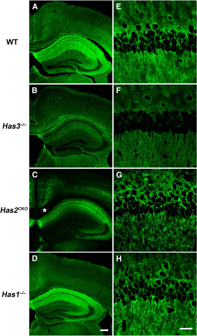
Spatial patterns of HA reduction in three Has mutant mouse models. Tissue staining of HA with biotinylated hyaluronan-binding protein (Laurent et al., 1991) was performed in the forebrain sections of WT (A, E), Has3−/− (B, F), Has2CKO (C, G), and Has1−/− (D, H) mice. Note that in WT mice, HA is strongly expressed in the hippocampus (A). In Has3−/− mice, hippocampal HA staining is markedly reduced (B). On the other hand, HA staining in the hippocampus is largely preserved in Has2CKO mice (C). Instead, reduction in HA is prominent in the superficial layers of the cortex and in fiber tracts in Has2CKO mice (e.g., the corpus callosum and the cingulum bundle; C, asterisk). The HA staining pattern in Has1−/− mice is essentially indistinguishable from that of WT mice (D). Scale bar, 200 μm. E–H, High-magnification images of the CA1 region. Note that not only is the level of HA staining greatly reduced in Has3−/− mice, but intercellular HA staining between neurons in the stratum pyramidale is essentially lost in Has3−/− mice (F). Scale bar, 30 μm.


