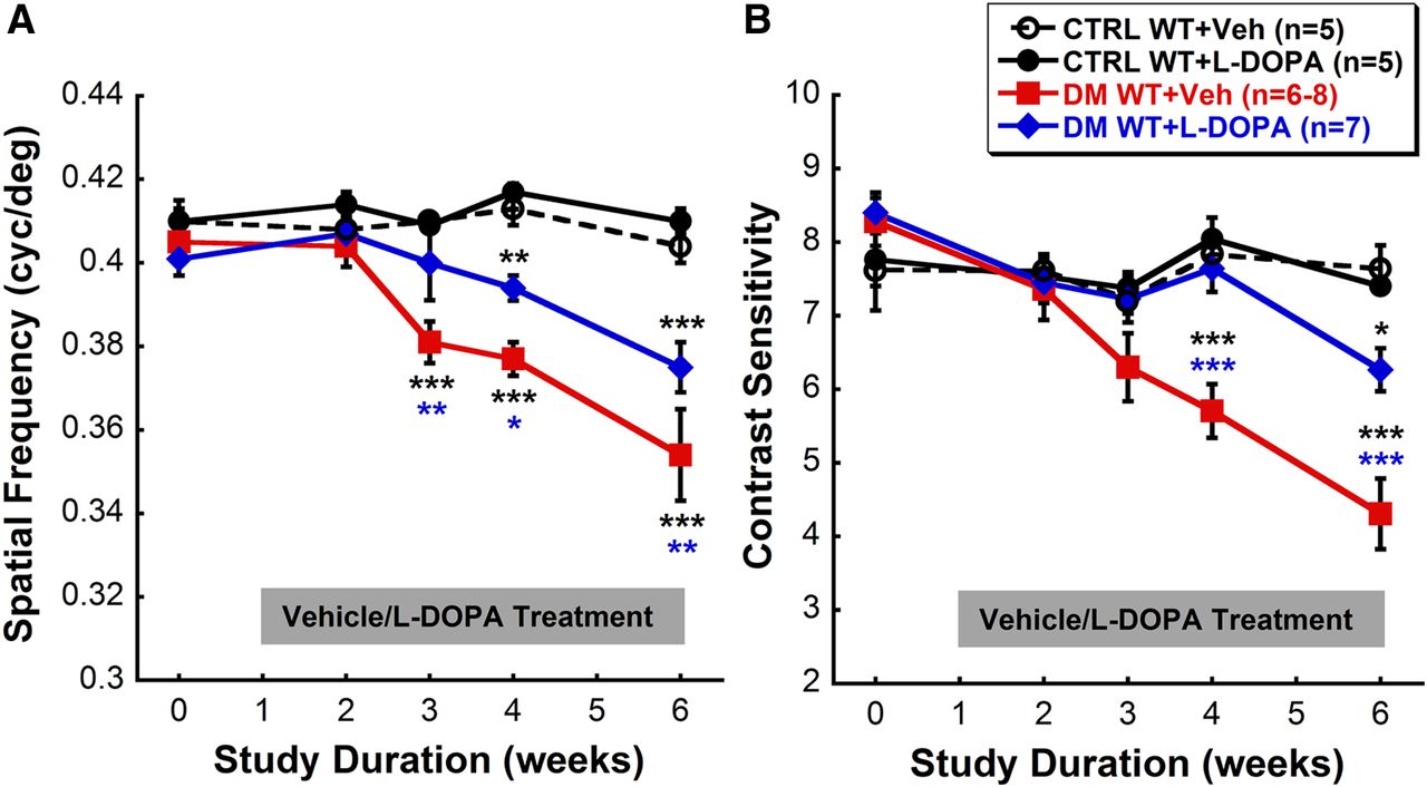
Chronic l-DOPA treatment delays early diabetes-induced visual dysfunction. A, DM WT + Veh mice showed significant reductions in spatial frequency threshold from CTRL animals as early as 3 weeks after STZ (post hoc comparison, p < 0.001). In contrast, the visual deficits appeared in DM WT + l-DOPA mice starting at 4 weeks after STZ (post hoc comparison, p < 0.01) with a slower and less severe progression. B, Contrast sensitivities were also significantly reduced in DM WT + Veh mice (post hoc comparison, p < 0.001) at 4 weeks after STZ, whereas DM WT + l-DOPA mice only exhibited a slight decrease in sensitivity at 6 weeks after STZ (post hoc comparison, p < 0.05). The color of the asterisk indicates the treatment group for which significance was reached, with the exception of the black asterisk, which refers to both CTRL WT + Veh and CTRL WT + l-DOPA groups. Note that the y-axis does not start at zero; it is modified to more clearly show the differences between treatment groups.


