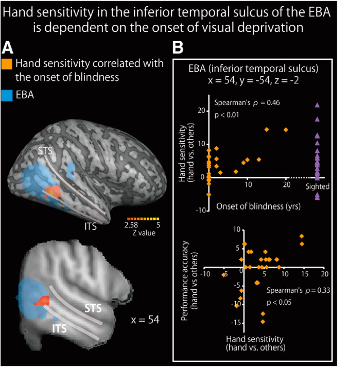
Hand sensitivity in the inferior temporal sulcus of the EBA is dependent on the age of visual loss (nonparametric analysis). A, Brain regions where hand sensitivity positively correlated with the age of visual loss are shown on a surface-rendered MRI (top row) and a sagittal section (bottom row) of an MRI averaged across the subjects. The activation was thresholded at p < 0.05, corrected for multiple comparisons in the EBA in each hemisphere, with the height threshold set at Z > 2.58. STS, Superior temporal sulcus; ITS, inferior temporal sulcus. See Table 8 for more information. B, The age at onset of blindness significantly predicted hand sensitivity (top) and hand sensitivity predicted performance accuracy (hands vs other objects) (bottom). Purple triangles represent the hand sensitivity of the sighted subjects. Data are presented as the mean ± SEM of 28 blind and 28 sighted subjects.


