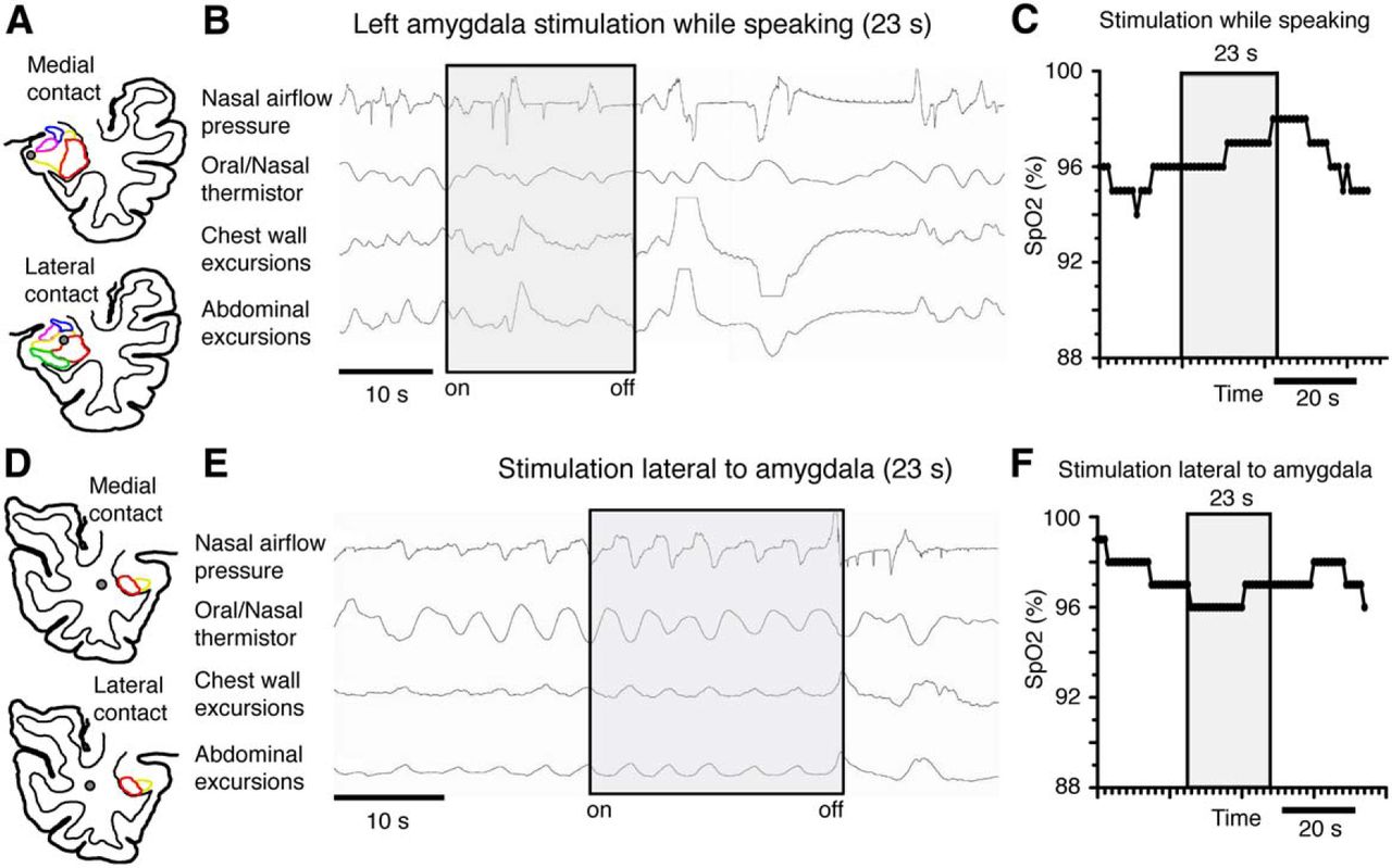
Apnea did not occur while speaking during amygdala stimulation and it did not occur during stimulation outside and lateral to the amygdala. A, Anatomic diagram of J.K.'s left temporal lobe showed amygdala subnuclei with superimposed amygdala depth electrode contacts (gray circles) in the basolateral (yellow) and lateral nuclei (red). B, C, Stimulating these contacts while speaking did not result in apnea and oxygen saturation (SpO2) remained unchanged, D, Anatomic diagram of J.K.'s right temporal lobe showing amygdala subnuclei with superimposed locations of depth electrode contacts (gray circles) immediately lateral to the amygdala. Stimulating these contacts just lateral and outside the amygdala did not cause changes in breathing (E) or oxygen saturation (SpO2; F). The nonspecific change in the plethysmography tracing immediately after stimulation (E) was associated with the patient's movement and when the patient stopped moving, the tracing returned to baseline.


