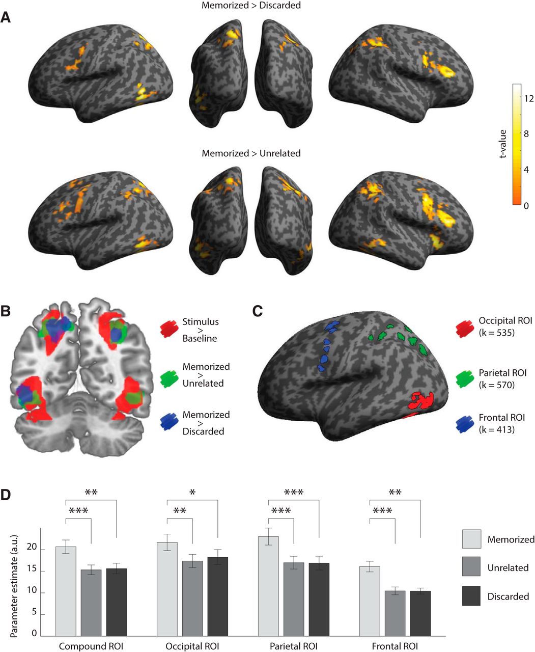
Results of the univariate analyses of BOLD response to the probe. A, Clusters of significant voxels (pcFWE < 0.05, for cluster-defining voxel threshold puncorrected < 0.001) from two contrasts, projected on an inflated surface of the standard Montreal Neurological Institute (MNI) template brain. None of the other contrasts (i.e., unrelated > discarded; discarded > unrelated; unrelated > memorized; and discarded > memorized) yielded significant clusters of voxels. B, A coronal slice encompassing visual processing areas in the occipital and parietal cortices (y-coordinate, 35 mm, MNI) on which the significant clusters of voxels from A are binarized and superimposed (80% opacity) on the significant clusters of voxels from the stimulus > baseline contrast of the functional localizer run (at the same voxel threshold, for illustrative purposes). C, Depiction of our ROIs. These ROIs comprised the significant voxels (p < 0.05FWE) in the stimulus > baseline contrast of the functional localizer run, separated into occipital (red), parietal (green), and frontal voxels (blue). D, Average parameter estimates for each Congruence condition (memorized, unrelated, discarded) against baseline, for each of the ROIs, as well as for the compound ROI comprising the occipital, parietal, and frontal ROIs together. Error bars represent the SEM. *p < 0.05, **p < 0.005, ***p < 0.0005.


