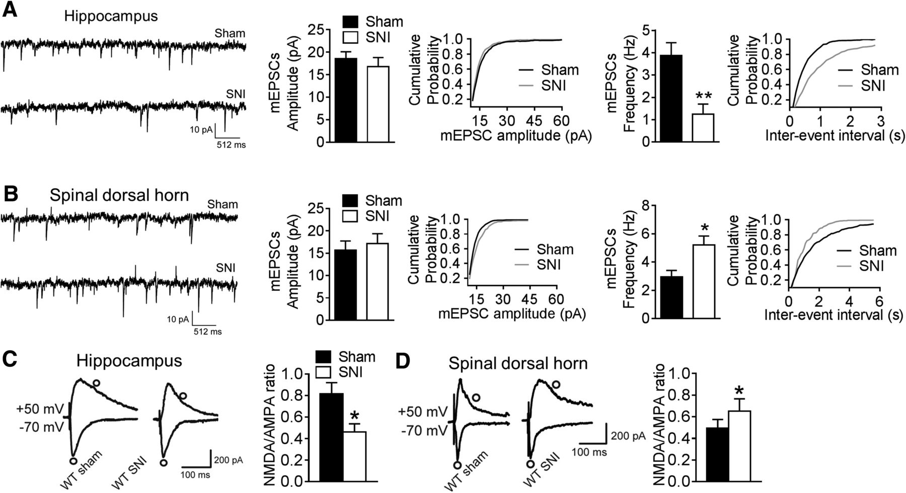
SNI induces differential changes in synaptic transmission. A, Representative mEPSCs of CA1 pyramidal neurons, their amplitude, cumulative probability, and frequency in WT mice from the sham and SNI groups (n = 12, amplitude, p = 0.659; frequency, p = 0.008; 12 neurons from 6 mice per group). B, Representative mEPSCs of spinal NK1-PNs, their amplitude, cumulative probability, and frequency in WT mice from the sham and SNI groups (n = 10, amplitude, p = 0.819; frequency, p = 0.012; 10 neurons from 5 mice per group). C, NMDA/AMPA current ratios at CA3–CA1 synapses in the indicated groups (n = 15, p = 0.021; 15 neurons from 6 mice per group). Evoked AMPA receptor EPSC (bottom traces) and evoked NMDA receptor EPSC (top traces) were recorded at membrane potentials of −70 and +50 mV, respectively. Circles indicate the time at which the amplitudes of AMPA or NMDA receptor currents were measured. D, NMDA/AMPA current ratio in spinal NK1-PNs in the different groups (n = 16, p = 0.022; 16 neurons from 6 mice per group). Values are the mean ± SEM. *p < 0.05, **p < 0.01 versus sham groups. Data for all bar graphs were analyzed using Student's t test.


