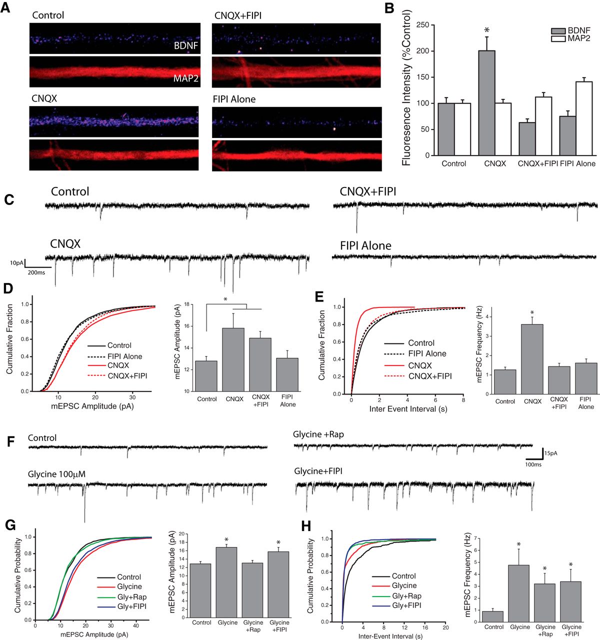
PLD signaling is required for adaptive presynaptic compensation. A, B, Examples images of BDNF and MAP2 expression in linearized dendritic segments (A), and mean (+SEM) expression (B) of MAP2 and BDNF in dendritic compartments normalized to average control values, in neurons treated with CNQX (40 mm, 3Hr) with or without the PLD1/2 inhibitor FIPI (100 nm, 30 min pretreatment). Control, n = 63; cells; CNQX, n = 75 cells; CNQX+FIPI, n = 70 cells; FIPI Alone, n = 56 cells. BDNF expression in dendrites was significantly enhanced by AMPAR blockade compared with control neurons. This effect was completely blocked by concurrent inhibition of PLD signaling. (*p < 0.05, relative to controls). C–E, Representative recordings (C) and mean (+SEM) mEPSC amplitude (D) and frequency (E) recorded from cells treated with vehicle control or CNQX (40 μm, 3Hr) with or without the PLD1/2 inhibitor FIPI (100 nm, 30 m pretreatment). Summary graphs presented on right and cumulative probability distribution shown on left. (*p < 0.05 relative to vehicle controls). Control, n = 22; CNQX, n = 9; CNQX+FIPI, n = 14; FIPI Alone, n = 18. PLD is required for the mTORC1-dependent (Henry et al., 2012) increases in mEPSC frequency observed after blockade of excitatory synaptic input. F–H, Representative recordings (F), and mean (+SEM) mEPSC amplitude (G) and frequency (H) recorded from cells treated with vehicle control or Glycine-based cLTP stimulus (100 μm glycine) with or without the PLD1/2 inhibitor FIPI (100 nm, 30 m pretreatment) *p < 0.05 relative to vehicle controls. Control, n = 12; Glycine, n = 11; Glycine+Rap, n = 8; Glycine+FIPI, n = 11. p < 0.05 compared with control. PLD signaling is not necessary for the mTORC1-dependent changes in mPESC Amplitude observed during chemically-induced LTP.


