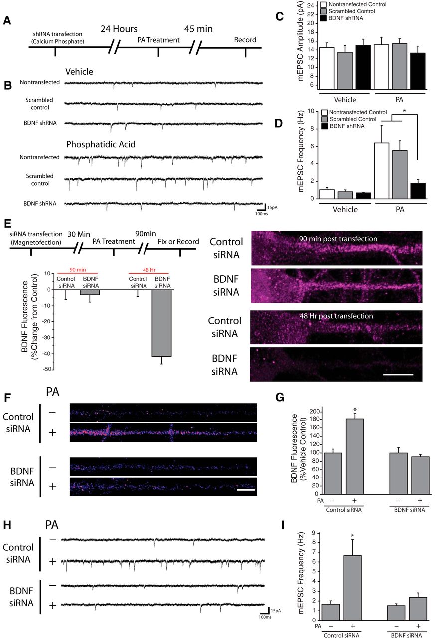
Postsynaptic BDNF expression is necessary for PA-induced changes in synaptic function. A, Experimental timeline: Dissociated hippocampal cultures were transfected with shRNA against BDNF or scrambled control via calcium phosphate. 24 h posttransfection, cells were treated with 100 μm PA for 45 min before recording. B–D, Representative recordings (B) and mean (+SEM) mEPSC amplitude (C) and frequency (D) in control nontransfected neurons (n = 7), as well as cells expressing BDNF shRNA (n = 8), or scrambled control shRNA (n = 4). The top panel in (B) contains representative traces from vehicle-treated group, while the bottom contains traces from cells subject to treatment with PA (100 μm, 45 min; n = 6, 9, 3). Cells were transfected at DIV 15 then used for recordings 24 h later. Postsynaptic BDNF knockdown blocked the increase in mEPSC frequency observed after PA treatment *p < 0.05 versus nontransfected cells and neurons transfected with scrambled shRNA. E, Experimental timeline: Dissociated hippocampal cultures were transfected with siRNA against BDNF or nontargeting control via magnetofection. 30 min posttransfection, cells were treated with phosphatidic acid for 90 min then either used for whole-cell recordings or fixed and processed for subsequent immunocytochemistry. (right) Representative images of straightened somadendritic regions of cultured neurons subject to magnetofection with control or anti BDNF siRNA. Scale bar represents 10 mm. Mean (+ SEM) expression of BDNF, normalized to average control values at identical time points, in neurons treated as indicated. While BDNF fluorescence was unaffected by magnetofection of anti-BDNF siRNA at 90 min (n = 21) compared with control siRNA transfected cells (n = 33), BDNF levels were significantly (*p < 0.05, relative to control) reduced by anti-BDNF siRNA magnetofection after 48 Hrs (n = 21) relative to neurons transfected with control siRNA (n = 32). F, G, BDNF expression in linearized dendritic segments, and mean (+ SEM) expression BDNF in dendritic compartments normalized to average control values, in neurons subject to magnetofection plus or minus PA (100 μm, 90 min). In neurons transfected with control siRNA, treatment with PA (n = 62) significantly enhanced BDNF expression in dendrites (*p < 0.05) compared with vehicle-treated cells (n = 55). In neurons transfected with BDNF siRNA, PA application (n = 60) failed to alter dendritic BDNF expression relative to vehicle (n = 49). Scale bar (in F): 10 μm. H, I, Representative recordings and mean (+ SEM) mEPSC frequency (H) in neurons subject to magnetofection plus or minus PA (100 μm, 90 min). Treatment with PA significantly enhanced mEPSC frequency in neurons transfected with the nontargeting control siRNA (*p < 0.05, t test; n = 6) compared with vehicle-treated neurons (n = 8). In neurons transfected with BDNF siRNA, no differences were observed between cells treated with PA (n = 7) or vehicle (n = 5).


