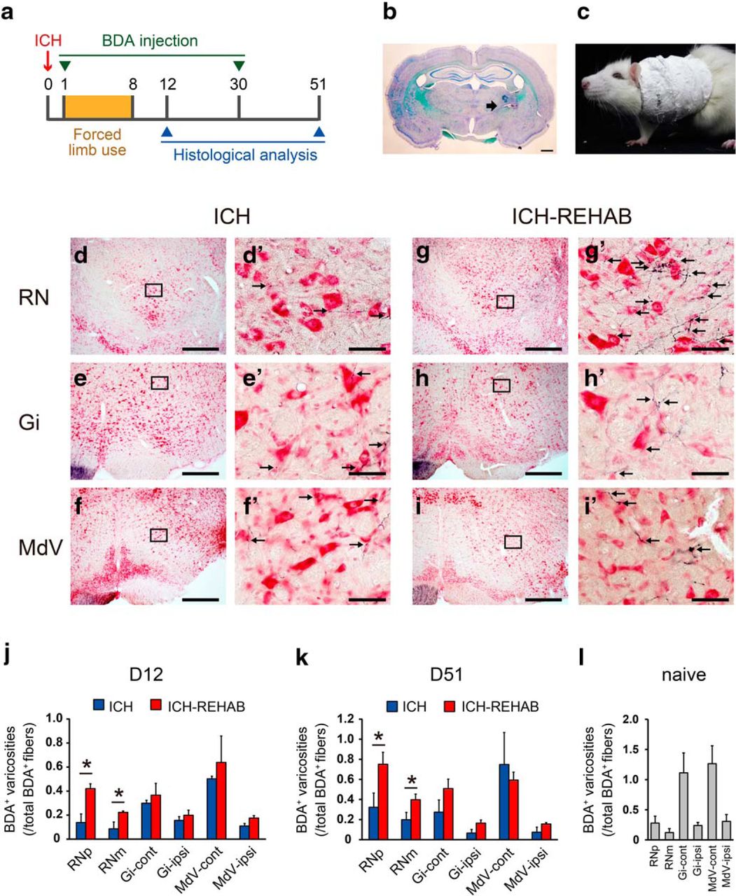
Axonal projections from the forelimb area of the ipsilesional motor cortex to the brainstem in the early and chronic phases of after ICH recovery. a, Timeline of the experiments. Forced use of the impaired forelimb was applied on postoperative days 1–8. BDA was injected into the ipsilesional motor cortex on after ICH day 1 or 30, and histological observation was conducted on day 12 or day 51. b, Representative Klüver–Barrera-stained image of an ICH. Arrow indicates the hemorrhage site induced by collagenase injection. c, Application of forced limb use treatment. A plaster cast restrained the unimpaired forelimb forcing the animal to use the impaired limb in their daily activities. d–i, Representative low-magnification images of BDA-labeled fibers in the major brainstem nuclei on day 12. The sections were counterstained with Neutral Red. d′–i′, Higher-magnification images of the squared areas in d–i. Arrows indicate varicosities apparently contacting neurons. d, g, Red nucleus (RN). Compared with the ICH group (d, d′), many BDA-positive fibers were found in the ICH-REHAB group (g, g′). e, h, Gi. f, i, MdV. j, k, Quantitative analysis for the varicosities of the cortico-rubral and cortico-reticular axons on day 12 (j) and day 51 (k). The number of BDA-positive varicosities in the parvocellular and magnocellular part of the red nucleus (RNp and RNm) and ipsilateral and contralateral Gi and MdV normalized against the total number of labeled axons in the cerebral peduncle is presented. Data for the number of BDA-positive varicosities in RNp and RNm at day 51 (k) are quoted from Ishida et al. (2016). The number of BDA-positive varicosities was significantly higher in the RN in the ICH-REHAB group (D12: n = 6; D51: n = 7) than in the ICH group (D12: n = 6; D51: n = 5), but not in reticular nuclei (Gi and MDV). l, The number of BDA-positive varicosities touching the red nucleus and the reticular nuclei in naive rats (n = 4). *p < 0.05. Data are mean ± SEM. Scale bars: b, 1000 μm; d–i, 500 μm; d′–i′, 50 μm.


