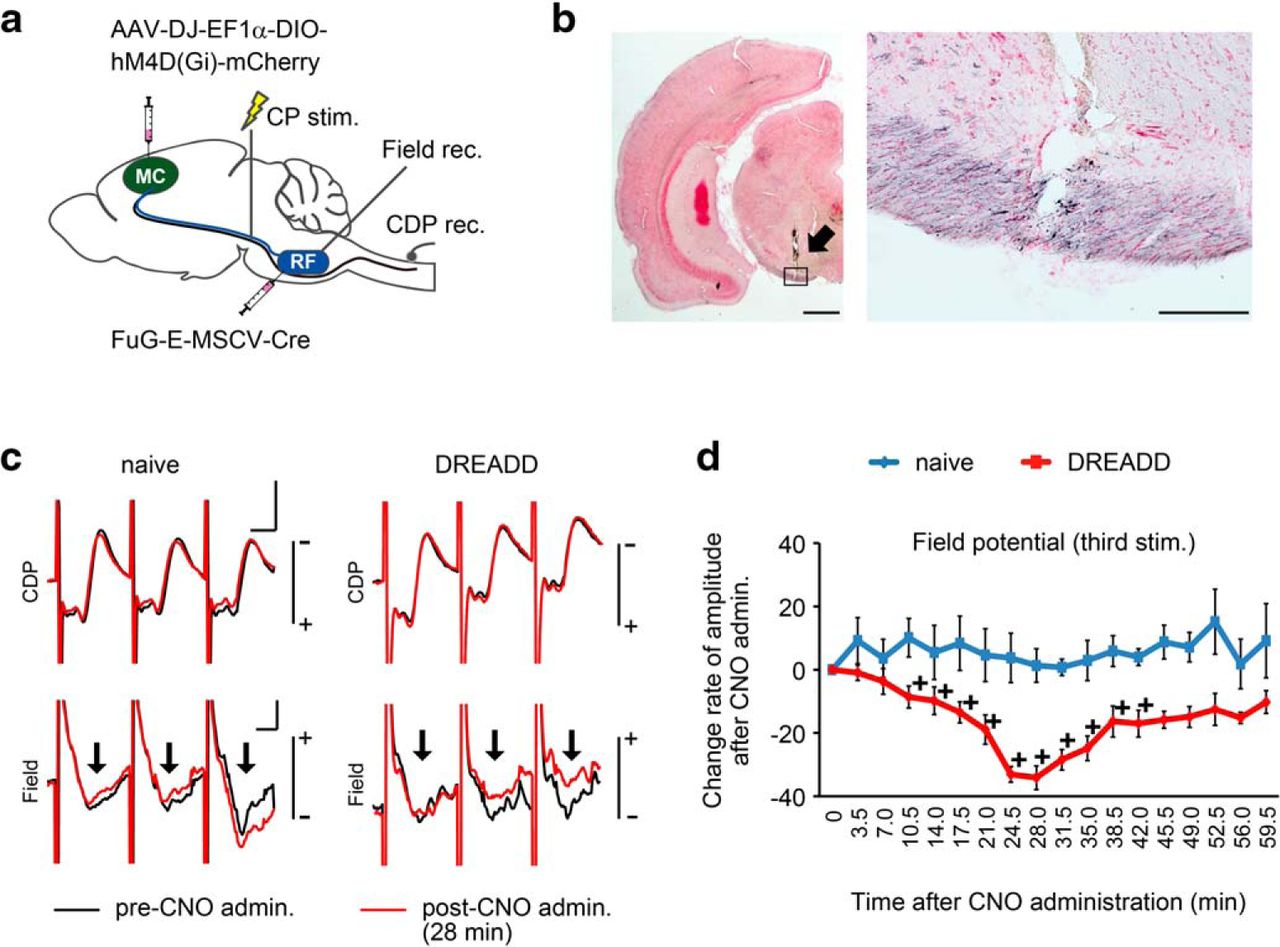
Electrophysiological confirmation of silencing of the cortico-reticular pathway by DREADD. a, Depiction of the experiment. Intact animals were injected with AAV-DJ-EF1α-DIO-hM4D(Gi)-mCherry into the forelimb area of the ipsilesional motor cortex (MC), and FuG-E-MSCV-Cre was injected into the caudal reticular formation (RF) bilaterally to disturb the cortico-reticular tract. To confirm successful silencing, field potentials (Field) in the MdV and CDP evoked by the stimulation of cerebral peduncle (CP) were recorded under anesthesia. b, Anti-mCherry immunohistochemistry of the stimulation site. Arrow indicates the trace of the electrode. Right, Higher magnification of the rectangle in left panel. The stimulation electrode reached the cerebral peduncle containing mCherry-positive fibers. c, Representative field potentials and CDP in a naive rat and in a rat with DREADD-induced silencing at before (black line) and 28 min after CNO administration (red line). Arrows indicate field potentials. Calibration: vertical bar, 1.0 mV for CDP and 0.2 mV for field; horizontal bar, 1 ms). d, Temporal alteration of LFP after CNO administration in naive-treated (n = 4) and DREADD-treated rats (n = 8). LFP decreased continuously in the DREADD-treated group from 10 to 40 min after CNO administration, and then recovered. +p < 0.05 versus 0 min after CNO administration. Data are mean ± SEM. Scale bars: b, Left, 1000 μm; b, Right, 200 μm.


