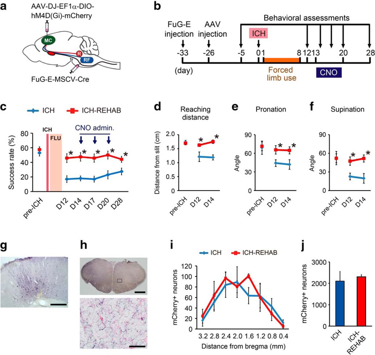
Effects of selective silencing of the cortico-reticular pathway on reaching function in the early-recovery phase. a, Depiction of the experiment. Animals were injected with AAV-DJ-EF1α-DIO-hM4D(Gi)-mCherry into the motor cortex (MC) and with FuG-E-MSCV-Cre into the bilateral reticular formation (RF) to silence the cortico-reticular tract neurons. b, Experimental time course. Animals were injected with viral vectors before ICH, and administered CNO on days 14, 17, and 20. Behavioral assessments were performed 5 d before ICH and 12, 14, 17, 20, and 28 d after ICH. Each test was conducted within 20–40 min after CNO administration. c, Averaged reaching success rate of ICH (n = 5) and ICH-REHAB groups (n = 6). The ICH-REHAB group showed significant recovery compared with the ICH group on day 12. Their performance was not affected by CNO administration. d–f, Detailed analyses of reaching movements. No obvious change was observed in reaching distance (d) or in the angle of wrist pronation (e) and supination (f). g, h, Anti-mCherry immunohistochemistry. mCherry-positive neurons were found in the ipsilesional motor cortex (g), and mCherry-labeled fibers were prominent in the contralateral caudal reticular nuclei (h). h, Bottom, Higher-magnification view of the top. i, Distribution of mCherry-labeled neurons in ICH (n = 4) and ICH-REHAB (n = 4) groups along the rostrocaudal axis. j, Averaged total number of mCherry-positive neurons of both groups. *p < 0.05 versus ICH. Data are mean ± SEM. Scale bars: h, Top, 1000 μm; g, 500 μm; h, Bottom, 100.


