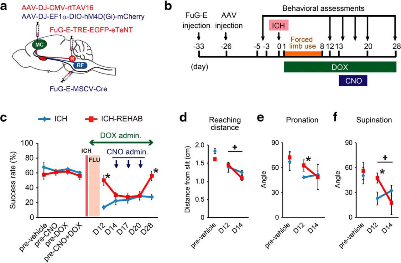
Effects of silencing the cortico-reticular pathway on functional recovery under continuous blockade of the cortico-rubral pathway from the start of rehabilitation. a, Experimental design. A mixture of AAV-DJ-CMV-rtTAV16 and AAV-DJ-EF1α-DIO-hM4D(Gi)-mCherry was injected into the ipsilesional motor cortex (MC) and FuG-E-TRE-EGFP-eTeNT and FuG-E-MSCV-Cre were injected into the ipsilateral red nucleus (R) and bilateral reticular formation (RF), respectively, to disturb the function of the cortico-rubral tract and the cortico-reticular tract (cortico-rubral tract) independently. b, Experimental time course. Two sets of FuG-E and AAV vectors were injected before ICH surgery. Behavioral assessments were performed before ICH and 12, 14, 17, 20, and 28 d after ICH. DOX was administered on days 1–28, and CNO was injected on days 14, 17, and 20. c, Average reaching success rate of ICH (n = 4) and ICH-REHAB groups (n = 6). The recovered function of the ICH-REHAB group showed significant impairment in response to CNO administration under the continuous administration of DOX. d–f, Average reaching distance (d) and the range of wrist pronation (e) and supination (f) in the skilled reaching test. Reaching distance and supination angle were significantly decreased in response to CNO administration in DOX-treated ICH-REHAB group rats. *p < 0.05 versus ICH. +p < 0.05. Data are mean ± SEM.


