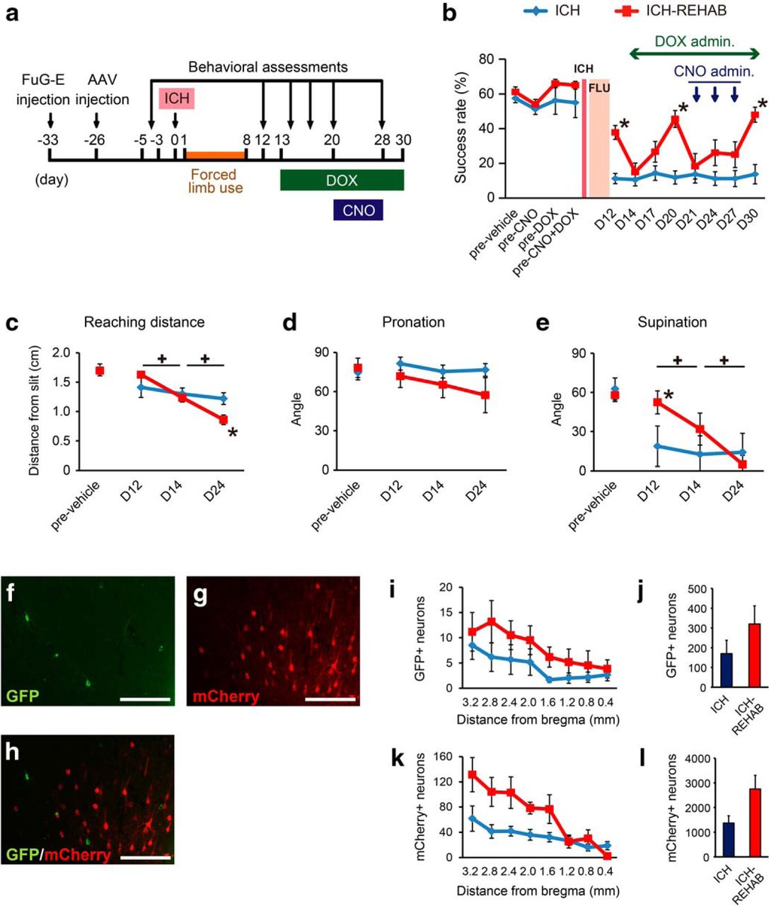
Confirmation of cortico-reticular pathway recruitment in response to blockade of the cortico-rubral pathway after stroke and rehabilitation. a, Experimental time course. Animals were injected with two sets of viral vectors before ICH surgery, as shown in Figure 6. Reaching performances were evaluated before ICH to provide a baseline and 12, 14, 17, 20, 21, 24, 27, and 30 d after ICH. DOX administration was conducted on days 13–30, and CNO was injected on days 21, 24, and 27. b, Average reaching success rate of ICH (n = 8) and ICH-REHAB groups (n = 9). The success rate of the ICH-REHAB group fell to the same level as the ICH group after DOX administration but recovered gradually. Additional injection of CNO caused a drastic decrease of the success rate. c–e, Qualitative analyses of the averaged reaching distance (c) and the range of wrist pronation (d) and supination (e). DOX administration caused a significant decrease of reaching distance and supination angle, and additional CNO administration decreased these abilities further. f–h, Representative images of GFP-positive (f) and mCherry-positive neurons (g) in the ipsilesional motor cortex. Few neurons expressed both GFP and mCherry (h). i, k, Distribution of GFP- and mCherry-labeled neurons along the rostrocaudal axis in the ICH (n = 6) and ICH-REHAB (n = 6) groups. j, l, Averaged GFP-positive (i) and mCherry-positive neurons (j) of both groups. *p < 0.05 versus ICH. +p < 0.05. Data are mean ± SEM. Scale bar, 100 μm.


