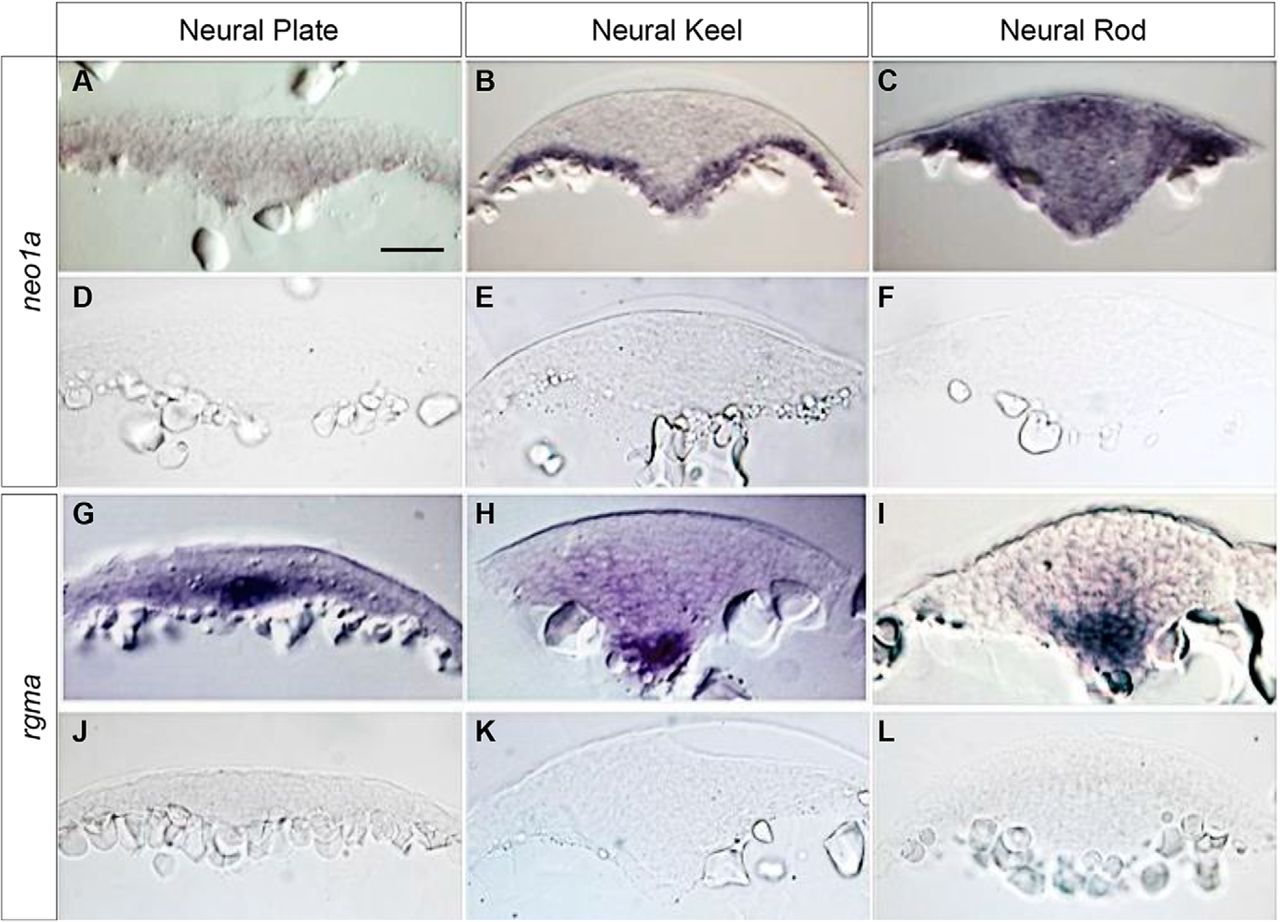
Figure 4.
Spatial distribution of zebrafish rgma and neo1a transcripts during NT development. Cross sections at the hindbrain level of zebrafish embryos at the NP (tailbud; A, D,G, J), neural keel (4–5 som; B, E, H, K), and neural rod (8–10 som; C, F, I, L) stages labeled by WISH using an antisense neo1a riboprobe (A–C), a sense neo1a riboprobe (D–F), an rgma antisense riboprobe (G–I), and a sense rgma riboprobe (J–L). Scale bar, 50 μm.


