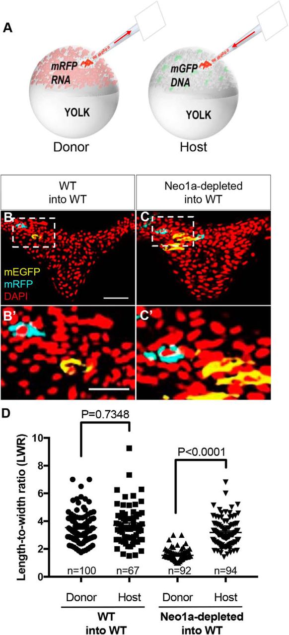
Neo1a functions cell autonomously in the neuroepithelium. A, Schematic representation of transplantation to produce chimeras. Cells were taken from the animal pole of shield stage donor embryos labeled with mRFP RNA and transplanted into the animal pole of host embryos at the same developmental stage that were mosaically labeled with mGFP DNA. Red arrows indicate the direction of donor cell movement through the transplantation needle. The host embryos were fixed at 4–5 som and analyzed for NEC morphology. B, C, Transverse sections through the hindbrain of 4–5 som chimeric embryos. mGFP and mRFP-expressing cells were immunolabeled with anti-GFP (yellow) and anti-RFP (cyan) antibodies, respectively. Nuclei were counterstained with DAPI (red). Scale bar, 50 μm. B′–C′, Insets, Higher magnifications of the selected regions in B and C. Scale bar, 25 μm. D, Quantification of the LWR of donor cells from uninjected controls (3.54 ± 0.1 μm, n = 100 cells from 18 embryos) and Neo1a-depleted embryos (1.54 ± 0.1, n = 92 cells from 9 embryos) and that of their control hosts (3.70 ± 0.2, n = 67 cells from 14 embryos and 3.18 ± 0.1, n = 94 cells from 10 embryos, respectively). Data are mean ± SEM.


