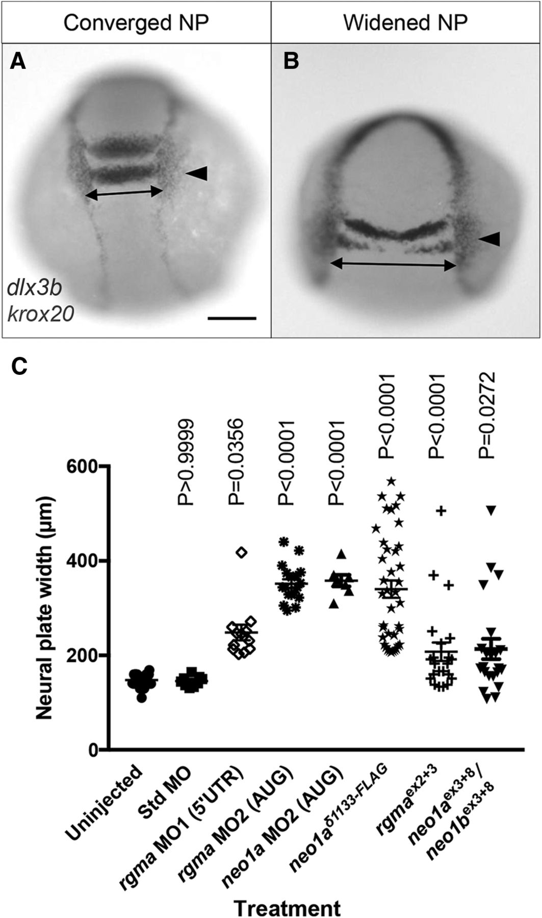
Figure 6.
Perturbation of Rgma/Neo1 signaling impairs NP infolding. A, B, WISH using dlx3 and krox20 probes on 4–5 som embryos that underwent timely (A) or delayed development (B). Arrow indicates lateral edge of krox20-positive rhombomere 5, which coincides with the position of the dlx3b-positive otic placode. Dotted line indicates NP width measured at the level of rhombomere 5/otic placode. Scale bar, ∼75 μm. C, Measurements of NP width (as indicated in A, B) of uninjected controls and embryos injected with std MO, rgma MO1, rgma MO2, neo1a MO2, neo1aδ1133-FLAG, rgma gRNA exon 2 + 3 and HF-nCas9 (rgmaex2+3), and neo1a/b gRNA exon 3 + 8 and HF-nCas9 (neo1aex3+8/neo1bex3+8).


