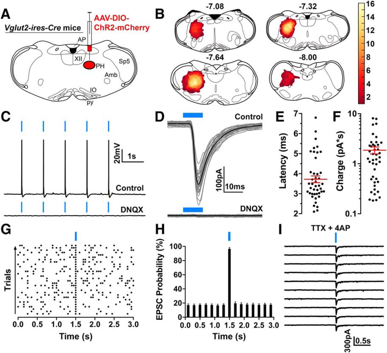
Photostimulation of parahypoglossal glutamatergic neurons activates hypoglossal motoneurons (PHGlut → HMNs). A, A schematic diagram of the experimental design we used to study synaptic connectivity between PH glutamatergic neurons and HMNs (PHGlut → HMNs). We injected an AAV-DIO-ChR2-mCherry into the PH region of VGlut2-IRES-Cre mice to express ChR2-mCherry in glutamatergic neurons. Three weeks after the AAV injections, we recorded in brain slices and photostimulated PHGlut input while recording from HMNs. B, Heatmaps of AAV-DIO-ChR2-mCherry injection sites of 16 recorded VGlut2-IRES-Cre mice. The yellow color indicates a higher number of overlapping regions of ChR2-mCherry-expressing neurons. C, Photostimulation of ChR2-expressing PHGlut neurons and terminals evoked action potential firing of HMNs (top trace), and this effect was blocked by the AMPA receptor blocker DNQX (20 μm; bottom trace), demonstrating the release of glutamate. D, In voltage-clamp mode, photostimulation of the PHGlut → HMN input evoked short-latency glutamatergic AMPA-mediated EPSCs in HMNs (in gray, 30 photo-evoked EPSCs; in black, average photo-evoked EPSC). E, F, Photo-evoked EPSC latencies (E) and charges (F) of photo-evoked EPSCs. Black dots, mean values of individual HMNs (average of 30 photostimulation trials); red lines, mean ± SEM, n = 43; average photo-evoked EPSC latency: 3.77 ± 0.17 ms, average photo-evoked EPSC charge: 1.86 ± 0.30 pA*s (or pC). G, H, A representative raster plot (G; 50 ms bins) and average EPSC probability (H) upon photostimulation of the PHGlut input (98.33 ± 1.44%; n = 43) and that of baseline without photostimulation (17.24 ± 1.62%; n = 43). I, Photo-evoked EPSCs were maintained in the presence of TTX (2 μm) + 4AP (1 mm), indicating monosynaptic connectivity. Voltage-clamp recordings were held at Vh = −60 mV. In all the recordings, we used 10 ms blue-light pulses, indicated by blue bars at the top of the recording traces. Atlas levels are per Paxinos and Franklin (2001).


