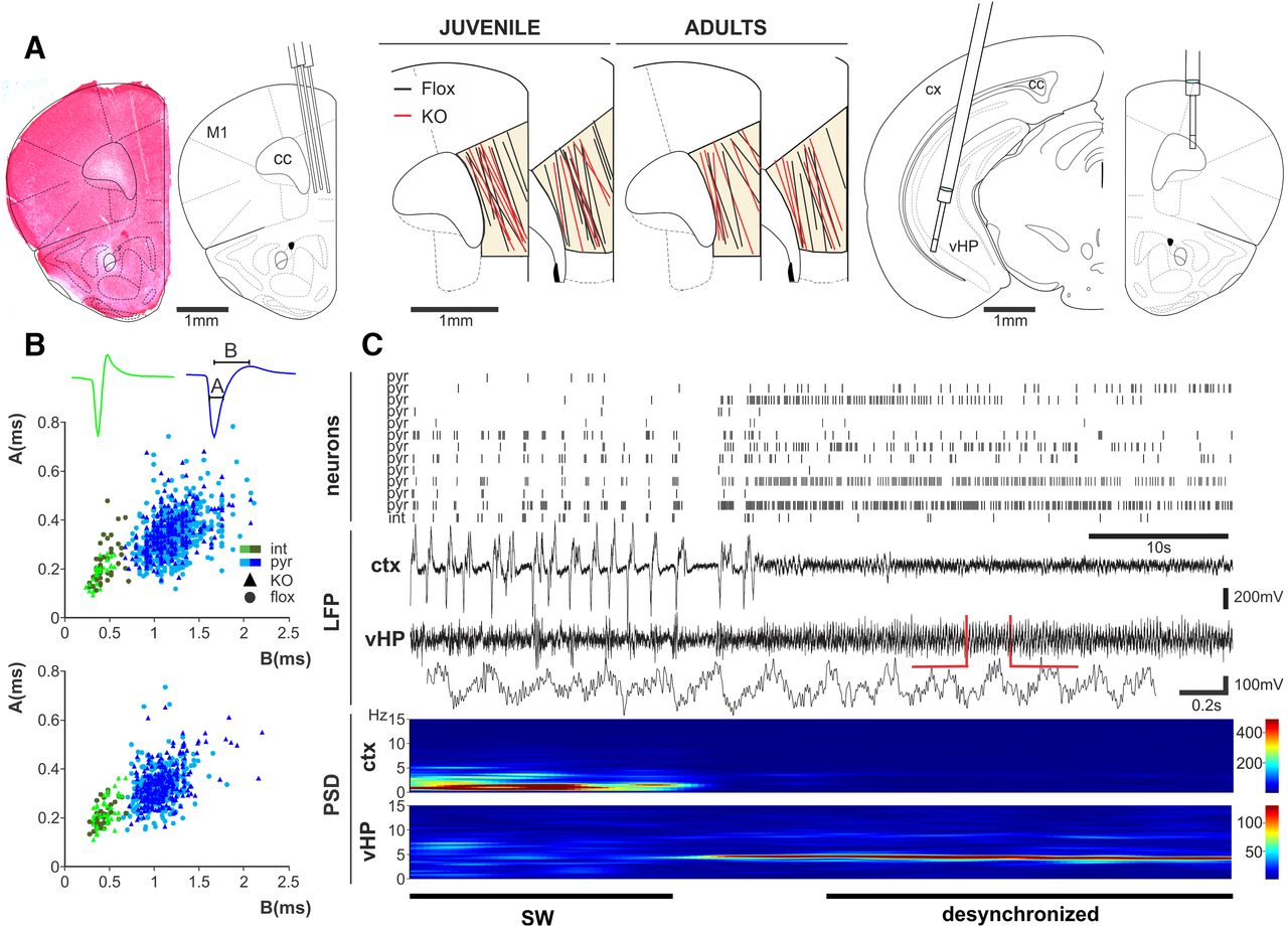
Simultaneously recording of unit activity in mPFC and LFP in cortex and hippocampus allows separation of putative neocortical interneurons and pyramidal cells during different global brain states. A, Left, Safranin O staining shows an example of the electrode track left by a tetrode recording in the mPFC, and schematic of a coronal section shows tetrode positioning in the mPFC. Middle, Schematic of coronal sections showing all electrode tracks in mPFC in juvenile (left) and adult (right) mice. mPFC recordings were performed along mPFC dorsoventral axis. No differences in electrode track positioning were observed across control (Flox, black lines) and KO (red lines) mice. Right, Schematic of coronal sections showing the localization of the concentric bipolar electrodes used to record the vHP LFP (left) and the cortical field potential (right). M1, Motor cortex; cc, corpus callosum; cx, cortex. B, mPFC pyramidal cell and interneuron classification based on their electrophysiological properties. Average waveforms of isolated units classified as interneuron (green) and pyramidal (blue), including temporal parameters used in the classification process (A, half-amplitude duration; B, trough to peak time). The parameters A and B were plotted for each cell, and two clear clusters were formed and used to sort neurons into pyramidal and interneurons in juvenile (top) and adult mice (bottom). C, Top, Representative raster plot of mPFC neurons (pyramidal and interneurons) recorded simultaneously with the cortical (ctx) and hippocampal (vHP) LFP activity. Bottom, Power density analysis performed on the same signal showing slow wave (SW) activity in the cortical LFP and a spontaneous desynchronization that correlates with the emergence of theta oscillations in the vHP.


