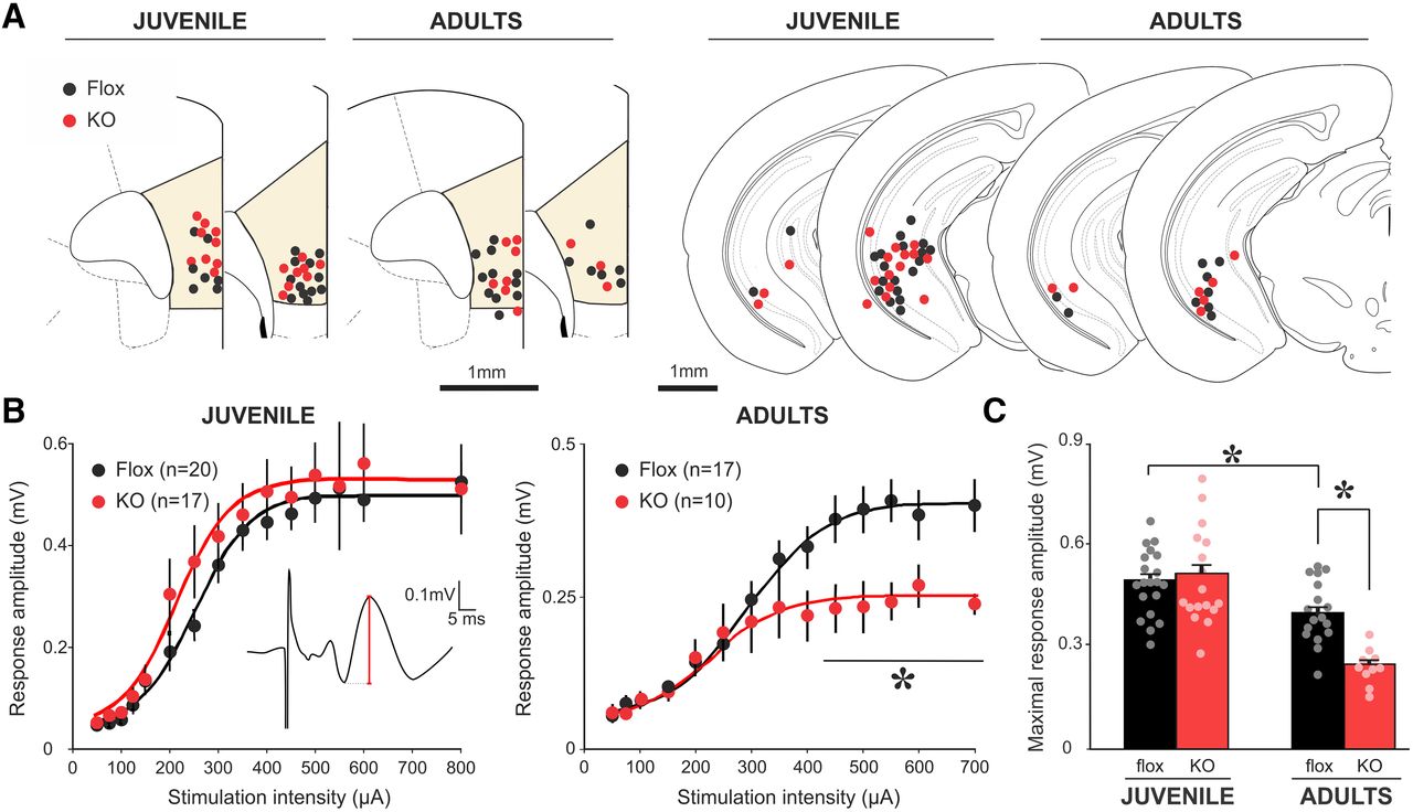
Only adult NMDAR-KO mice show a reduced mPFC-evoked response to vHP stimulation. A, Schematic of coronal sections showing recording (mPFC; left) and stimulation (vHP; right) electrode positioning for juvenile and adult control and KO mice. B, Stimulation of vHP evoked a short-latency response in mPFC LFP (inset; red line indicates amplitude of the evoked response). mPFC stimulus-response curves evoked by stimulation of vHP show a significant decrease in response amplitude in adult, but not juvenile, KO mice compared with age-matched control mice (two-way repeated-measures ANOVAs). Adult: genotype × intensity interaction, p < 0.001. *p < 0.05 (Newman-Keuls post hoc test vs control at same stimulation intensity). Juvenile: genotype factor, p = 0.49; genotype × intensity interaction, p = 0.62. Each point represents mean ± SEM. Number of mice is indicated. Trend line indicates the best fit sigmoidal curves to population data. C, Maximum evoked mPFC response to vHP stimulation. Adult KO mice show a decreased maximal evoked response compared with age-matched control mice, although no differences were observed between control and KO juvenile mice (two-way ANOVA, genotype × age interaction). *p < 0.05 (Newman-Keuls post hoc test). Error bar indicates mean ± SEM. Each dot represents 1 individual animal.


