This Week in The Journal
- Teresa Esch, Ph.D.
Control of Pectoral Fin Movements in Zebrafish
Yuto Uemura, Kagayaki Kato, Koichi Kawakami, Yukiko Kimura, Yoichi Oda, et al.
(see pages 6678–6690)
Locomotion is produced by alternating contraction of antagonist muscles within limbs or on opposite sides of the body. The timing and sequencing of these contractions are controlled by central pattern generators (CPGs) that provide rhythmic input to motor neurons. The best understood locomotor CPGs in vertebrates are those of lamprey and tadpoles, which swim by body undulation. In these species, CPGs that drive contraction of trunk musculature are present in each segment of the spinal cord. Bursting of ipsilateral glutamatergic interneurons drives motor neurons, while commissural inhibitory interneurons ensure that contractions alternate between the two sides.
Swim CPGs were likely repurposed during evolution to control walking in limbed vertebrates. CPGs for walking must be more complex, however, because they coordinate movements of multiple joints both within and between limbs. Zebrafish offer an intermediate level of complexity. Although zebrafish swim by body undulation, their pectoral fins—homologous to forelimbs—move forward and back coordinately with this undulation, with left and right fins moving in opposite directions. These movements are less complex than walking, however, because fins lack joints. Therefore, Uemura et al. investigated CPGs controlling pectoral fin movements in zebrafish larvae.
The authors traced the motor neurons innervating adductor (medial) and abductor (lateral) fin muscles to the rostral spinal cord, where neurons innervating the dorsal-most fin muscles were located most rostrally. As expected, abductor and adductor motor neurons fired in alternating rhythmic bursts, each with a set phase relationship to bursts in motor neurons that innervated the body wall. During each phase of the swim cycle, fin motor neurons received tonic excitatory input as well as phasic excitatory and inhibitory input. Examination of the activity of several classes of interneurons defined by transcription factor expression revealed that commissural glycinergic interneurons expressing dmrt3a provide rhythmic inhibitory input to abductor motor neurons, ensuring proper timing of bursts during swimming.
Previous work indicated that dmrt3a neurons regulate alternating contraction of axial muscles during swimming. Notably, the axons of dmrt3a neurons in segments containing fin motor neurons were shorter than those in the rest of the trunk. This is consistent with the hypothesis that CPG neurons that control swimming are repurposed to control appendages. Future work will provide further elucidation of this repurposing.
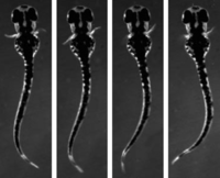
While larval zebrafish swim, their pectoral fins move in an alternating rhythm that is coordinated with movement of the body. See the article by Uemura et al. for details.
Development of the Extrinsic Innervation of the Gut
Xueyuan Niu, Li Liu, Tao Wang, Xin Chuan, Qi Yu, et al.
(see pages 6691–6708)
The gastrointestinal tract has its own nervous system—the enteric nervous system—with sensory, motor, and interneurons that detect gut contents, drive contractions, and regulate secretion, absorption, and local blood flow. But the gut also receives extrinsic innervation from sensory afferent fibers and sympathetic and parasympathetic efferent fibers. Although this extrinsic innervation is not required for the gut to function normally, it mediates voluntary control of gut input and output and enables the gut to communicate with the brain. For example, the fibers transmit information about satiety and inflammation from gut to brain, and about emotional and cognitive states from brain to gut.
During embryonic development, extrinsic afferent and efferent fibers reach the gut after intrinsic innervation has begun. Because afferent and efferent fibers often travel in the same nerves, however, determining the spatiotemporal pattern of growth of individual fiber classes has been difficult. Niu, Liu, et al. have now identified specific markers for each fiber type, and they used these markers to follow the development of extrinsic innervation of the gut.
Sensory fibers from the nodose ganglion extended in the vagus nerve and reached the esophagus by embryonic day 10.5 (E10.5). Parasympathetic axons arising in the dorsal motor nucleus in the medulla followed closely behind pioneer sensory axons in the vagus nerve, and these fibers reached the stomach by E11.5. Sympathetic fibers began to extend toward the gut at E12.5, and then grew along the gut mesentery, where they were joined by growing vagal sensory and parasympathetic axons. The three types of axons traveled together, bifurcated into gastric and celiac branches, and reached the rostral hindgut by E14.5. Notably, ablating vagal sensory neurons impaired targeting of vagal parasympathetic axons, as well as delaying innervation of the gut by sympathetic fibers. Finally, as vagal and sympathetic fibers extended caudally along the gut, lumbosacral spinal sensory neurons extended from dorsal root ganglia, joined with caudal sympathetic and parasympathetic fibers, and innervated the hindgut. Thus, the entire surface of the gut was innervated by E16.5.
This study lays the groundwork for future investigations of the molecules guiding extrinsic innervation of the gut and how targeted disruption of this innervation affects gut–brain communication.




