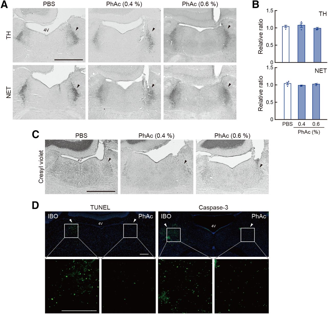
Morphologic analysis of LC neurons after PhAc stimulation. A, Histology of LC NE neurons in the Tg mice after PhAc treatment. Sections through the LC were prepared from the Tg mice used for microdialysis analysis 7 d after unilateral treatment with PBS or PhAc (0.4/0.6%) and stained for TH and NET immunohistochemistry. B, Cell counts for TH- and NET-immunopositive cells. The ratio of the number of cells stained for TH or NET in the treated side relative to the intact side was calculated. Data are presented as the mean ± SEM. Individual data points are overlaid. C, Staining of LC sections with cresyl violet, showing no cell damage against LC neurons in the PhAc-treated side. D, Staining for cell death markers. IBO (1 mg/ml) or PhAc (0.6%) was unilaterally injected into the LC (0.2 μl/site) of the Tg mice. LC sections were stained with TUNEL or immunohistochemistry for activated caspase-3. The LC areas in the top images were fourfold magnified in the lower images. Arrowheads indicate the injection sites into the LC. 4V, Fourth ventricle. Scale bars: A, C, D, 1 mm.


