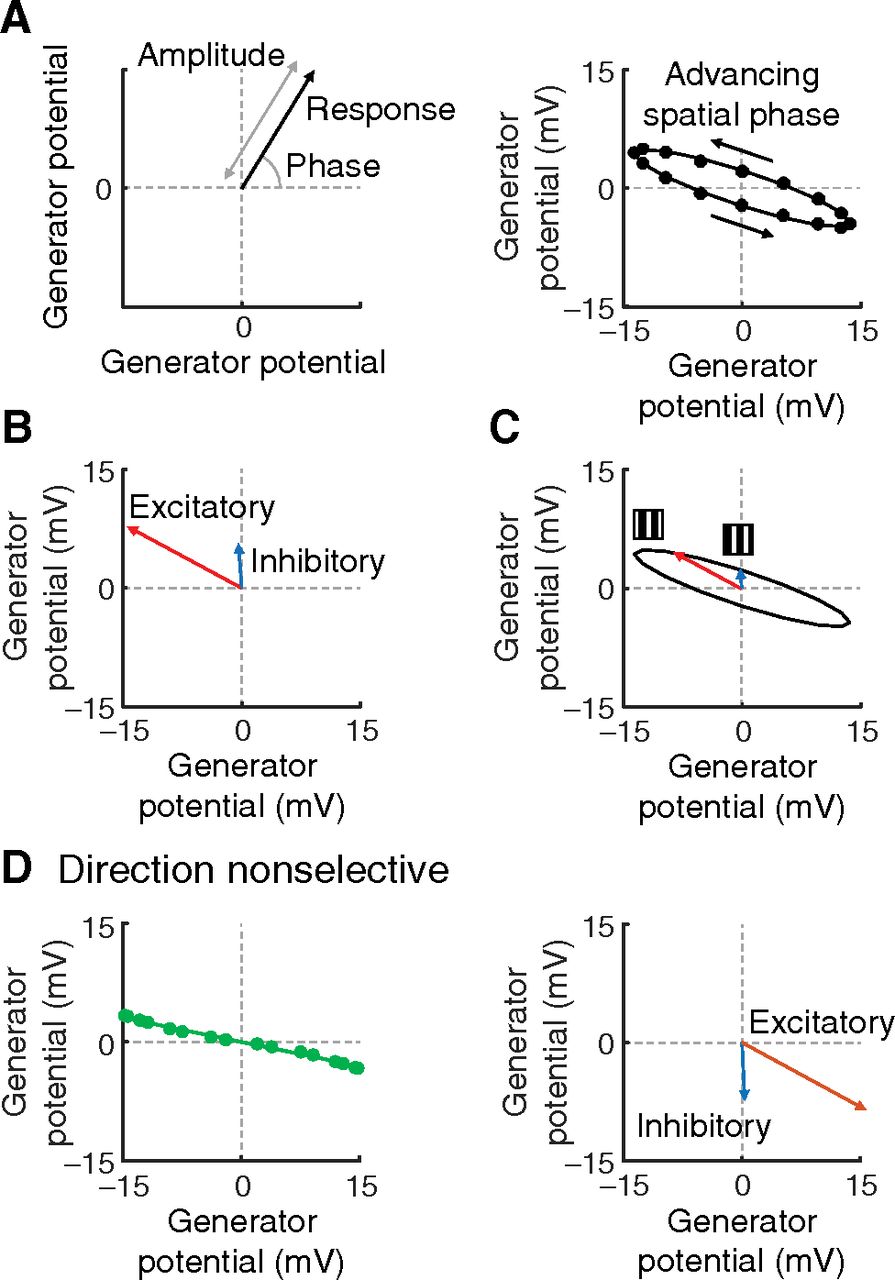
Mechanism of direction selectivity. A, Graphs in this figure use polar coordinates where the length and angle of a vector give, respectively, the amplitude and phase of the fundamental Fourier component of the response to a contrast-reversing grating. Right, Graph represents the result for the direction-selective neuron represented in Figures 3A, 9A, and 10A. The points shown by circles were calculated from the model, and the ellipse comes from vector summation as illustrated in B. Spatial phase advances anticlockwise around the ellipse. B, Both the excitatory and inhibitory inputs to an excitatory cell are insensitive to stimulus direction, have phase plots like those in Figure 9B (right), and are therefore well approximated by vectors with a fixed response phase. C, Their amplitudes, however, depend on spatial phase as shown here. Response phase advances with spatial phase because of the spatial phase offset of the inhibitory vector relative to the excitatory vector. D, Polar plots for the direction-nonselective cell in Figure 3A are shown for comparison.


