This Week in The Journal
- Teresa Esch, Ph.D.
Oxytocin Affects Development of Dopamine Neurons
Ana Rita Nunes, Michael Gliksberg, Susana A. M. Varela, Magda Teles, Einav Wircer, et al.
(see pages 8742–8760)
Oxytocin has important roles in social behaviors, such as pair-bonding, parenting, social affiliation, and aggression, in birds and mammals. It also regulates stress responses, depression-related behaviors, and sensory processing. Although most studies of oxytocin and its receptors have focused on acute effects in adult animals, oxytocin may also influence behavior by regulating the development of underlying circuitry, much like gonadal steroids shape neural circuits involved in adult reproductive behaviors. Indeed, Nunes, Gliksberg, et al. provide evidence for such an organizational effect of oxytocin in zebrafish.
Although zebrafish are gregarious animals that typically associate with other fish in shoals, previous work had suggested that oxytocin receptors play only a limited role in social interaction. Consistent with this, chemically ablating oxytocin neurons in adult zebrafish had no effect on the tendency of singly housed fish to spend time near a separate compartment containing conspecifics. Surprisingly, however, adult fish in which oxytocin neurons had been ablated at the larval stage showed a reduced tendency to stay near the shoal. Remarkably, this effect occurred only when oxytocin neurons had been ablated before social affiliation behavior had developed and occurred even though the neurons had regenerated by the time of testing.
In addition to altering social affiliation behavior in adults, larval ablation of oxytocin neurons led to a decrease in the number of dopaminergic neurons in the pretectum and posterior tuberculum within 24 h. Notably, unlike oxytocin neurons, dopamine neurons did not regenerate by adulthood. Finally, larval ablation of oxytocin neurons altered neuronal responses and activity patterns in several brain areas involved in social behavior and decision-making.
These results suggest that oxytocin is required for normal development of dopaminergic neurons in areas involved in visual processing (the pretectum, analogous to mammalian superior colliculus) and motivation/reward (the posterior tuberculum, analogous to the ventral tegmental area) in zebrafish. These effects might impair recognition of shoals and diminish the motivation to affiliate with shoals, both of which, along with altered activity in other brain areas, could explain the behavioral effects seen in adult fish. Future work should examine whether oxytocin has similar organizational effects in mammals.
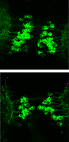
Adult fish in which oxytocin neurons were ablated during larval development (bottom) have fewer dopaminergic neurons (green) in the pretectal area than normal (top). See Nunes, Gliksberg, et al. for details.
GABAergic Neurons in Medial Amygdala Promote Lordosis
Caroline S. Johnson, Weizhe Hong, and Paul E. Micevych
(see pages 8790–8800)
When a female mouse in estrus encounters an attractive male, she will assume the lordosis posture—back arched, head and rear up, tail deflected—signaling her willingness to mate. This behavior is controlled by hypothalamic neurons, including those in the ventrolateral subdivision of the ventromedial hypothalamus (VMHvl) that project to brainstem regions controlling motor output. Information about the presence and attractiveness of a potential mate are conveyed to the hypothalamus partly by the posterodorsal portion of the medial amygdala (MeApd), which receives input from the pheromone-sensing vomeronasal organ, as well as other sensory areas. Because GABAergic MeApd neurons influence multiple social behaviors, Johnson et al. guessed they are also necessary for lordosis. Conversely, they hypothesized that glutamatergic MeApd neurons inhibit lordosis.
In separate groups of female mice, the authors expressed halorhodopsin selectively in GABAergic MeApd neurons or expressed channelrhodopsin selectively in glutamatergic MeApd neurons. They then measured lordosis in the presence and absence of optical stimulation when mice were placed with a sexually experienced male. Whereas inhibiting GABAergic neurons decreased lordosis, exciting glutamatergic neurons had no effect. Nevertheless, exciting glutamatergic MeApd neurons increased the time spent in self-grooming, which is considered an antisocial behavior. The authors also found that inhibiting GABAergic MeApd neurons reduced the number of VMHvl neurons that expressed the activity marker c-Fos, whereas activating glutamatergic neurons had no effect on c-Fos expression in hypothalamic nuclei. Finally, in control mice that exhibited normal levels of lordosis when placed with a male, c-Fos levels were increased more frequently in GABAergic MeApd neurons than in glutamatergic MeApd neurons.
These results support the hypothesis that GABAergic MeApd neurons promote lordosis. Notably, the fact that inhibiting these neurons reduced c-Fos expression in VMHvl suggests they exert their behavioral effect by increasing activity in VMHvl. In contrast, glutamatergic MeApd neurons do not directly inhibit lordosis, but instead increase the time spent engaged in activity not obviously related to reproduction. Future work should identify the specific neuronal populations targeted by GABAergic projections from MeApd to VMHvl and other brain areas involved in reproductive behaviors.




