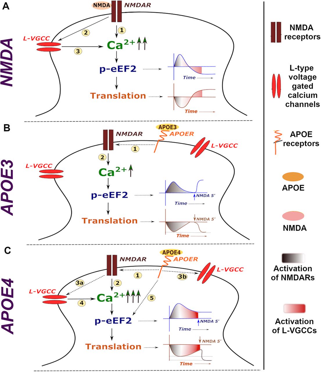
Model illustrating calcium signature and corresponding protein synthesis regulation downstream of NMDAR stimulation and APOE treatment. A, Stimulation of NMDARs lead to influx of calcium through NMDARs (1) which activates L-VGCCs (2). The influx of calcium through L-VGCCs further helps in sustaining the calcium levels on NMDAR stimulation (3). The calcium through NMDARs and L-VGCCs generates a specific temporal profile of eEF2 phosphorylation, causing an initial inhibition of global protein synthesis followed by translation activation in a later phase. B, Exposure to APOE3 activates NMDARs through an unknown mechanism (1), leading to a short burst of calcium through it (2). This leads to an acute increase in eEF2 phosphorylation, primarily contributed by calcium influx through NMDARs, which recovers to basal levels. Global translation also follows a similar temporal profile of initial decrease followed by recovery. Hence, the NMDA activity mediated translation response is unaffected in APOE3-treated neurons. C, Exposure to APOE4 activates NMDARs through an unknown mechanism (1), causing higher influx of calcium through NMDARs (2) than APOE3 condition. The higher calcium influx through NMDARs could lead to L-VGCC activation in APOE4 condition (3a). Besides, APOE4 binding to APOE receptors could also directly regulate the sustained activation of L-VGCCs (3b). Overall, the L-VGCC activation under APOE4 treatment condition contributes to the huge and sustained increase in calcium levels (4). This leads to the sustained increase in eEF2 phosphorylation as well as global translation inhibition. There could also be a possibility of APOE4 activating signaling cascades which further contribute to the sustained increase in eEF2 phosphorylation (5). Hence, the NMDA activity mediated response is perturbed, potentially causing a stress-response phenotype in APOE4-treated neurons.


