Understanding the Influence of Target Acquisition on Survival, Integration, and Phenotypic Maturation of Dopamine Neurons within Stem Cell-Derived Neural Grafts in a Parkinson's Disease Model
- Niamh Moriarty,
- Jessica A. Kauhausen,
- Chiara Pavan,
- Cameron P.J. Hunt,
- Isabelle R. de Luzy,
- Vanessa Penna,
- Charlotte M. Ermine,
- Lachlan H. Thompson, and
- Clare L. Parish
- Florey Institute of Neuroscience and Mental Health, The University of Melbourne, Parkville, Victoria 3010, Australia
-
Author contributions: C.L.P. and L.H.T. designed research; C.L.P., J.A.K., I.R.d.L., C.M.E., L.H.T., N.M., C.P.J.H., V.P., and C.P. performed research; C.L.P., J.A.K., I.R.d.L., C.M.E., L.H.T., N.M., C.P.J.H., V.P., and C.P. analyzed data; C.L.P. and N.M. wrote the first draft of the paper; C.L.P., J.A.K., I.R.d.L., C.M.E., L.H.T., N.M., C.P.J.H., V.P., and C.P. edited the paper.
Abstract
Midbrain dopaminergic (DA) neurons include many subtypes characterized by their location, connectivity and function. Surprisingly, mechanisms underpinning the specification of A9 neurons [responsible for motor function, including within ventral midbrain (VM) grafts for treating Parkinson's disease (PD)] over adjacent A10, remains largely speculated. We assessed the impact of synaptic targeting on survival, integration, and phenotype acquisition of dopaminergic neurons within VM grafts generated from fetal tissue or human pluripotent stem cells (PSCs). VM progenitors were grafted into female mice with 6OHDA-lesions of host midbrain dopamine neurons, with some animals also receiving intrastriatal quinolinic acid (QA) injections to ablate medium spiny neurons (MSN), the A9 neuron primary target. While loss of MSNs variably affected graft survival, it significantly reduced striatal yet increased cortical innervation. Consequently, grafts showed reduced A9 and increased A10 specification, with more DA neurons failing to mature into either subtype. These findings highlight the importance of target acquisition on DA subtype specification during development and repair.
SIGNIFICANCE STATEMENT Parish and colleagues highlight, in a rodent model of Parkinson's disease (PD), the importance of synaptic target acquisition in the survival, integration and phenotypic specification of grafted dopamine neurons derived from fetal tissue and human stem cells. Ablation of host striatal neurons resulted in reduced dopamine neuron survival within grafts, re-routing of dopamine fibers from striatal to alternate cortical targets and a consequential reduced specification of A9 dopamine neurons (the subpopulation critical for restoration of motor function) and increase in A10 DA neurons.
- dopamine
- human pluripotent stem cells
- Parkinson's disease
- synaptic plasticity
- target acquisition
- transplantation
Introduction
Development of the central nervous system is a highly complex process involving billions of axons forming functional connections with their appropriate targets. In early neural development, the first axons navigate through the largely axon-free environment, with their growth cones detecting and responding to guidance cues and interacting with the underlying extracellular environment to eventually form an initial target contact. These “pioneer axons” are thought to lay down a path on which subsequent axons follow (Harrison, 1959). This is supported by studies in embryonic zebrafish where the ablation of pioneer axons resulted in aberrant axon pathfinding (Chitnis and Kuwada, 1991). During development neurons are generated in excess, forming many more synaptic connections than are necessary for function, resulting in ∼50% undergoing cell death. This is thought to occur in an attempt to fully and adequately innervate the target (Oppenheim, 1991). Quantitatively, cell death is most dramatic when neurons start forming synaptic connections to their target cells suggesting that target acquisition plays a role in survival and maturation (Marti et al., 1997; Dekkers et al., 2013). In support, partial deletion of targets typically leads to reduced survival while increasing the availability of targets reduces cell death (Hamburger et al., 1981; Oppenheim, 1991; Dekkers et al., 2013). This is in contrast to the adult brain where ablation of target cells bares no impact on the survival of afferent neurons, inferring that mature neurons remain less dependent on target-derived support (Sofroniew et al., 1990; Lundberg et al., 1994).
During rodent midbrain DA development, cell death occurs within the first two postnatal weeks (Oo and Burke, 1997). The death of these DA neurons is thought to be regulated by an interaction with their target medium spiny neurons (MSNs) within the striatum as striatal lesioning (Macaya et al., 1994), DA terminal destruction (Marti et al., 1997) and axotomy of the medial forebrain bundle (El-Khodor and Burke, 2002) all result in reduced numbers of DA neurons. This has been supported by in vitro studies demonstrating that ventral midbrain (VM) tissue co-cultured with their target neurons promoted their development (Prochiantz et al., 1979) and survival (Hoffmann et al., 1983).
Target acquisition has also been shown to influence DA neurons within VM fetal grafts -transplants aimed at restoring dopamine transmission and function in Parkinson's disease (PD). Studies have observed greater DA survival and plasticity when VM progenitors are implanted into a striatum devoid of host afferent dopaminergic connection compared with grafts placed into an intact striatum (Björklund et al., 1983; Doucet et al., 1990; Zuddas et al., 1991). These findings were assumed to be the result of target availability as grafts preferentially connected with the denervated areas of the striatum (Björklund et al., 1983). What remains less known is the specific influence of the postsynaptic target cell on the survival and plasticity of DA grafts.
There is also a lack of knowledge on the influence that the target neuron has on the maturation and fate acquisition of subpopulations of DA neurons both in development and within VM progenitor grafts. Within the developing VM, and VM-derived grafts, reside two predominant subpopulations of DA neurons, the A9 neurons of the substantia nigra pars compacta that can be identified by the expression of G-protein-gated inwardly rectifying K+ channel subunit 2 (GIRK2) and project via the nigrostriatal pathway to the dorsolateral striatum to regulate motor function, and the A10 DA neurons that co-express Calbindin, and project via the mesocorticolimbic pathway to cortical and limbic structures to regulate reward-related behavior (Dahlstrom and Fuxe, 1964; Mendez et al., 2005; Thompson et al., 2005). In PD, these A9 DA neurons are the first to degenerate and contribute to the various motor anomalies observed in patients (Agid et al., 1993) and unsurprisingly are the population necessary for functional recovery following grafting into preclinical PD models (Mendez et al., 2005; Kuan et al., 2007; O'Keeffe et al., 2008; Grealish et al., 2010). Despite their importance, our knowledge of what influences the specification of A9 DA neurons, over adjacent A10 neurons, remains surprisingly limited. In light of the role of target acquisition in survival and plasticity of VM DA neurons in development and neural transplants we hypothesized additional roles of the postsynaptic cell in DA fate acquisition. Here, we demonstrate in a Parkinsonian model, that ablation of the striatal MSN has a significant influence on the integration of both rodent fetal and human pluripotent stem cell (PSC)-derived VM DA progenitors, affecting graft survival, regional plasticity, and DA fate acquisition.
Materials and Methods
Animals
All animal procedures adhered to the Australian National Health and Medical Research Council (NHMRC)'s Code of Practice for the Use of Animals in Research and were approved by the Florey Institute of Neuroscience and Mental Health animal ethics committee. Animals were group housed in individually ventilated cages on a 12:12 h light/dark cycle with ad libitum access to food and water. A total of 38 adult female Swiss mice were used as graft recipients in transplantation study involving mouse fetal tissue, and 16 adult female athymic (Foxn1nu) nude mice were used as hosts for human PSC-derived VM progenitor grafts.
Preparation of VM donor cells
Fetal VM donor tissue was isolated from embryonic day (E)12.5 mice that expressed green fluorescent protein under the tyrosine hydroxylase promotor (TH-GFP; Sawamoto et al., 2001). The tissue was enzymatically dissociated into single cell suspensions using previously described methods (Kauhausen et al., 2013). The VM progenitors were resuspended at a density of 100,000 cells/µl in magnesium and calcium-free HBSS with the addition of DNase (0.1%). Cell suspensions were stored on ice until the time of grafting. Note, the use of the GFP reporters (driven by the TH) enabled clear distinction of graft (from host) dopamine neurons and their axonal processes.
Generation of hiPSC PITX3-eGFP reporter line and VM progenitors
The human-induced PSC (hiPSC) line, RM3.5, was maintained on Laminin-521 (0.5 µg/cm2, Biolamina) as previously described (Gantner et al., 2020a). To generate the novel reporter line, engineered to express GFP under the PITX3 promoter. Dissociated hiPSCs were collected in resuspension buffer R (Neon 100 µl kit, Invitrogen) and 1 µg/1.0 × 106 cells of PITX3-eGFP-PGK-PURO donor plasmid (originally a gift from Rudolf Jaenisch, Addgene deposit #31942; from the PITX3 CompoZr Targeted integration kit (Sigma) were incubated on ice for 5 min. Following incubation, equimolar ratios of PITX3 zinc-finger nuclease plasmids (ZFN1 and ZFN2) were added and the sample transferred to the Neon (Invitrogen) pipette using the Neon tip. Cells were electroporated under optimised conditions (1650 V, 10-ms pulse width and a pulse number of 3) and replated in hiPSC media (at 1:3, 1:6, 1:9 dilutions).
Approximately 72 h following electroporation, Puromycin (0.5–1.0 µg, Thermofisher) was added for ∼11 d to select for successful donor plasmid integration. Puromycin-treated colonies (from 1:9 dilution) were manually dissected and expanded for an additional 6 d. Following expansion, cells were isolated and genomic DNA (gDNA) was extracted using the ISOLATE II genomic isolation kit (Bioline). Insertion of donor construct at PITX3 locus was confirmed by Phusion high-fidelity RT-PCR kit (ThermoFisher). In brief, 20 ng gDNA (1 ng for vector control), 3% DMSO and all remaining kit reagents were combined with in-house designed primers to amplify through the left homology arm of the donor plasmid. cDNA samples were run on a two-step amplification protocol using the Veriti thermal cycler (ThermoFisher) programmed for 30 s at 98°C for initial denaturation, 35 cycles of 10 s at 95°C for denaturation, 90 s at 72°C for extension, and a final extension at 72°C for 10 min. Following thermocycling PCR samples were mixed with 6× TriTrack loading dye (ThermoFisher) and electrophoretically separated at 90 V for 45 min on a 1% (w/v) agarose gel in 1 × TAE buffer alongside HyperLadder™1 kb ladder (Bioline) with relevant controls and visualized at 340 nm using the UV imaging system (BioDoc-It). Successful integrated clones displayed bands at 984 bp, while vector-only or human gDNA controls gave rise to bands at 1350 and 970 bp, respectively.
Human VM progenitors were generated from the PITX3-GFP hiPSC line and differentiated as previously described (Gantner et al., 2020a), with cells collected for transplantation at day 19. The resultant VM progenitors were dissociated using Accutase (Innovative Cell Technologies) and resuspended at a density of 100,000 cells/µl in NBB27 media with ROCK inhibitor (Y27632, 10 µm). The cell suspension was stored on ice until the time of grafting.
Surgical procedures
All animal surgeries were performed under general anesthetic using 2–5% isoflurane inhalation (Baxter). A single unilateral injection of 6OHDA (3.2 µg dissolved in 0.02% ascorbic acid in sterile saline) was delivered into the substantia nigra at the following coordinates in Swiss (and nude) mice: 3.2 mm (3.0 mm) posterior, and 1.2 mm (1.0 mm) relative to bregma and 4.5 mm (4.2 mm) below the surface of the dura. During the same surgery, a proportion of 6OHDA-lesioned mice also received an injection of quinolinic acid (QA; Sigma-Aldrich; 60 nmol in 0.5 µl) into the ipsilateral, dorsolateral striatum (1.0 mm anterior and 2.0 mm lateral to bregma, and 3.0 mm below the dura surface). At three weeks after lesioning, mice received intrastriatal injections (1 µl) of mouse fetal-derived or hiPSC-derived VM progenitors (coordinates: 1.0 mm anterior and 2.0 mm lateral to bregma, and 3.0 mm below the dura surface).
Tissue processing and immunohistochemistry
For confirmation of appropriate in vitro specification of VM neural progenitors, cultures were fixed using paraformaldehyde (PFA; 4%, 10 min) on day 13 and 30 of differentiation. For assessment of in vivo graft survival, animals were terminally anaesthetized and perfused 12 weeks after transplantation. Animals received a lethal overdose of sodium pentobarbitone (100 mg/kg, Virbac) and were transcardially perfused with 60–80 ml of warmed (37°C) PBS followed by 30 ml of chilled PFA (4% w/v). Fixed brains were carefully removed and postfixed in the same fixative for 2 h before being transferred to a sucrose solution (20% w/v in 0.1 m PBS) overnight. When the brains reached equilibrium (sunk within the sucrose solution) they were serially cut into 40-µm sections (1:12 series) using a freezing microtome (Leica).
Free-floating sections were quenched in 0.3% H2O2/methanol in PBS for 20 min, washed (PBS; 3 × 5 min) and incubated overnight in the primary antibody diluted in 5% donkey serum and 0.3% Triton X-100 (Ameresco). For fluorescent staining, no H2O2 quenching was required, and tissue sections (or in vitro cultures) were washed (3 × 5 min in PBS) and directly incubated in primary antibodies. Primary antibodies were as follows: mouse anti-Nestin (1:1000, R&D Systems), goat anti-FOXA2 (1:200, Santa Cruz), goat anti-OTX2 (1:500, R&D Systems), rabbit anti-NURR1 (1:200, Santa Cruz), rabbit anti-TH (1:800, Pel-freeze), sheep anti-TH (1:800, Pel-freeze), rabbit anti-BARHL1 (1:200, Novus Biologicals), sheep anti-PITX2 (1:200, R&D Systems), rabbit anti-GFP (1:200, Abcam), chicken anti-GFP (1:1000, Abcam), mouse anti-NeuN (1:200, Millipore), rabbit anti-DARPP32 (1:200, Abcam), rabbit anti-GFAP (1:200, DAKO), mouse anti-CD11b (1:100, Serotec), mouse anti-synaptophysin (1:1000, human specific, Enzo Life Sciences), mouse anti-calbindin (1:1000, Swant), rabbit anti-GIRK2 (1:500, Abcam), goat anti-GIRK2 (1:500, Abcam), and rabbit anti-ALDH1A1 (1:400, Abcam). The following day, the sections (or cultures) were washed in PBS before being incubated in secondary antibody (diluted in 2% donkey serum and 0.3% Triton X-100) for 2 h. Tissue was washed with PBS and chromogenically stained sections incubated in avidin–biotin peroxidase for 1 h (Vectastain ABC kit, Vector Laboratories) before the final peroxidase reaction using 3,3′-diaminobenzidine tetrahydrochloride solution (DAB; Sigma-Aldrich). The secondary antibody used for DAB-based detection was biotin-conjugated donkey anti-rabbit (1:200, Jackson ImmunoResearch). For direct fluorescent detection DyLight 488-, 549-, or 649-conjugated donkey anti-mouse, anti-chicken, anti-goat, or anti-rabbit antibodies (Jackson ImmunoResearch; 1:200) were used. Fluorescently labeled sections were mounted on to gelatin-coated slides, and coverslipped using fluorescent mounting media (DAKO). Chromogenically stained tissue was dehydrated in a series of alcohol baths and xylene before being coverslipped using DePex mounting media (BDH Chemicals).
Imaging and quantification
All fluorescent images were captured using a Zeiss LSM780 confocal system or a Zeiss Axio ObserverZ.1 upright epifluorescence microscope. Brightfield images were taken on a Leica DM6000 upright microscope, and when required stitched using LAS (Leica) software. For GFP+ cell counts, cells were counted from live images at 20× magnification. Graft core, innervation areas and DARPP32 lesions were measured by delineating the areas of brain sections inhabited by GFP+ cells or fibers, or devoid of DARPP32 staining, respectively, and volumes estimated using Cavalieri's principles (Cavalieri, 1966). The immunologic response of the host brain to QA and 6OHDA lesioning was assessed at predetermined sites lateral to the graft-host border (delineated according to GFP labeling). The area covered by GFAP or CD11b immunoreactivity was expressed as a percentage of the total pixels in the intact, 6OHDA+QA, and 6OHDA-lesioned brain.
Innervation density was measured from 20× color inverted images by isolating GFP+ fibers using the “color range” tool on photoshop (Adobe) and expressing values as a percentage of total immunoreactive pixels. Assessment of GIRK2 and Calbindin expressing cells within grafts was quantified based on co-localization with GFP from acquired confocal images.
Transcriptional profiling
Transcriptional profiling, by quantitative real-time PCR (Q-PCR), was performed on maturing VM cultures derived from the PITX3-GFP IPSC cell lines. Cell pellets (at Day (D) 9, 13, 19, 22, 25, and 30) were lysed using a TissueLyser LT (QIAGEN) and total RNA extracted using the RNeasy Mini kit (QIAGEN). RNA yield and integrity were assessed using a Nanodrop One spectrophotometer (ThermoFisher Scientific) and confirmed using a Qubit (Thermo Fisher Scientific) and Tapestation (Agilent). Q-PCR was performed using the ABI7700 sequence detection system (Applied Biosystems), and data assessed using the comparative ΔΔCT method. See Table 1 for a list of primers.
List of primers used for Q-PCR
Statistical analysis
All data were expressed as the mean ± SEM. All statistical analyses were conducted using GraphPad Prism 5. Student's t tests or one-way ANOVA (with Bonferroni's multiple comparisons test) were used to identify differences within the data. Significance was set at p < 0.05.
Results
Absence of a striatal target does not influence the survival of fetal-derived ectopic transplants
To determine whether the availability of MSN targets influenced VM grafts, donor tissue (isolated from embryos expressing GFP under the TH promoter; TH-GFP) was ectopically transplanted into the striatum of 6OHDA-lesioned mice with or without a QA striatal lesion. Immunostaining against DARPP32 and NeuN confirmed that the vast majority (>65%) of striatal MSNs were lost in QA-lesioned animals within 10 d (Fig. 1A,B), resulting in significant striatal atrophy at 10 weeks (Fig. 1C; striatal volume in Intact: 12.57 ± 1.253 mm3, 6OHDA: 13.11 ± 1.153 mm3, 6OHDA+QA: 4.99 ± 0.595 mm3; p < 0.0001, Fig. 1H). Unsurprisingly at 10 d after QA lesion, during periods on ongoing MSN neuron degeneration, the host striatum showed significantly elevated density of GFAP+ reactive astrocytes (Fig. 1D,F) and CD11b microglia (Fig. 1E,G), compared with the intact brain. Animals receiving only 6OHDA lesions (in the absence of QA) also showed a significantly elevated astrocytic and microglial response at this time, a response to the ongoing degeneration of the DA terminal field (Fig. 1D–G), as previously reported (Stott and Barker, 2014). Despite these changes to the surrounding striatum, QA lesioning had no effect on the volume of the graft core, i.e., delineation of GFP+ cell bodies (6OHDA: 0.232 ± 0.044 mm3; 6OHDA+QA: 0.400 ± 0.070 mm3, respectively, p = 0.067; Fig. 1I,L,M) and no impact on the number of GFP+ DA neurons (6OHDA: 1313 ± 136; 6OHDA+QA: 1492 ± 230 GFP+ cells, p = 0.1888; Fig. 1J). Consequently, with no difference in graft core volume or GFP+ cell numbers, the density of DA neurons remained unchanged (6OHDA: 7.48 × 10³ ± 1.55 × 10³, 6OHDA+QA: 5.55 × 10³ ± 0.99 × 10³ GFP+ cells/mm3, p = 0.306; Fig. 1K).

Loss of striatal targets does not affect the survival of ectopically transplanted fetal-derived neurons. Representative images illustrating MSNs, identified by DARPP32+ and NeuN+ co-expression, within the striatum (A) that were ablated within 10 d of QA lesioning (B). Significant striatal atrophy was evident at 10 weeks in QA-lesioned animals (C, H). 6OHDA and 6OHDA+QA lesioning evoked an inflammatory response in the host striatum, reflected by a significant increase in GFAP+ reactive astrocytes (D) and, CD11b+ reactive microglia (E). Representative photomicrographs showing GFAP (F) and CD11b (G) labeling in the striatum of the intact, 6OHDA, and 6OHDA+QA-lesioned brain. The loss of a striatal target by QA lesioning did not influence the volume of fetal VM graft core (I, p = 0.083; delineated by the dotted white line; L', M'), GFP+ cell survival (J, p = 0.1888) or graft core density (K, p = 0.429). Photomicrographs illustrating placement of GFP+ fetal VM grafts into the 6OHDA and 6OHDA+QA-lesioned striatum, respectively (L, M). GFP+ labeling of representative A9-like and A10-like DA neurons (L'', M''). Data are mean ± SEM; ***p < 0.001, ****p < 0.0001 versus intact; ##p < 0.01, ###p < 0.001 versus 6OHDA. n = 4–8/group (D, F) n = 7–8/group (H), n = 18–19/group (I–K). Scale bars: 1 mm (A–C, L, M, L', M'), 500 µm (A'–C'), 100 µm (E–G, L''–M'').
Absence of a striatal target significantly impacts graft plasticity
We next assessed the impact the loss of striatal target cells had on the innervation and plasticity of VM fetal tissue grafts. Striatal reinnervation from the graft, defined as the volume of the striatum occupied by GFP+ fibers (Fig. 2A,B, white dashed lines), was significantly reduced in QA-lesioned animals compared with non-QA-lesioned controls (2.58 ± 0.27 and 5.373 ± 0.52 mm3, respectively; p < 0.0001; Fig. 2C). Furthermore, analysis of DA innervation density within the dorsolateral striatum (the primary target region of the striatum that underpins motor function) revealed a significant reduction in GFP+ (DA) fiber density (40.88 ± 2.48% vs 10.13 ± 1.26%, respectively; p < 0.0001; Fig. 2Ai,Bi,D). While the density of striatal innervation strongly correlated with GFP+ cells numbers (6OHDA: r2 = 0.60, p = 0.0001, open circles), this correlation was no longer present in QA-lesioned animals that lacked striatal targets (6OHDA+QA: r2 = 0.15, p = 0.1162; Fig. 2F, closed circles).

Target availability influences patterns of graft-derived GFP+ fiber innervation of the host brain. Representative darkfield images of graft-derived GFP+ innervation within host tissue (A, B, white line delineates GFP+ fibers within the confines of the striatum). Magnified regions illustrating GFP+ innervation in the striatum (Ai, Bi) and cortex (Aii, Bii) of 6OHDA and 6OHDA+QA-lesioned animals, respectively. Quantitative analysis revealed that QA lesioning significantly reduced both the volume (C, p < 0.0001) and density (D, p < 0.0001) of striatal innervation, while increasing the fiber density within the cortex (E, p < 0.0001). The strong correlation between the number of GFP+ neurons and striatal innervation observed in 6OHDA grafted animals was lost in 6OHDA+QA animals (F, 6OHDA: r2 = 0.60 vs QA: r2 = 0.15). In contrast, where 6OHDA+QA-lesioned animals showed an inverse correlation in striatal innervation relative to the % DARPP32 lesioning (G, r2 = 0.70; black dots), preferential innervation of the cortex was evident by the positive correlation between DARPP32 lesion size and density of GFP+ fibers in the cortex (G, r2 = 0.43; red dots). Data are mean ± SEM; ***p < 0.001, ****p < 0.0001. n = 18–19/group (C–F), n = 8/group (G). Scale bars: 1 mm (A, B) and 100 µm (Ai, Aii, Bi, Bii).
With a loss of striatal targets observed in QA-lesioned animals, GFP+ fiber density significantly increased in the overlying cortex, an additional target of VM DA neurons (6OHDA: 2.1 ± 0.71%; 6OHDA+QA: 14.33 ± 2.87% GFP+ pixels, p = 0.0005; Fig. 2Aii,Bii,E). This shift in regional targeting was most evidently reflected in comparative regression analysis, illustrating the inverse correlation between the degree of DARPP32 lesioning and the density of GFP fibers within the striatum (r2 = 0.70, p = 0.017; Fig. 2G, black line), and the positive correlation between DARPP32 lesioning and cortical innervation (r2 = 0.43, p = 0.109; Fig. 2G, red line).
Loss of striatal targeting influences the phenotype of grafted DA neurons
Within the mammalian VM, subsets of DA neurons show preferential target association. A9 DA neurons of the substantia nigra pars compacta, expressing GIRK2, preferentially synapse striatal neurons, while A10 DA neurons of the ventral tegmental area, expressing Calbindin, target limbic areas, including cortex. With a shift in graft innervation observed in QA-lesioned mice, we assessed the impact on DA phenotyping. Immunohistochemical staining against GFP, GIRK2 and Calbindin enabled evaluation of dopaminergic phenotype acquisition of grafted neurons in 6OHDA and 6OHDA+QA-lesioned mice (Fig. 3A–D). Reflective of our previous observations (Thompson et al., 2005; Bye et al., 2012; Kauhausen et al., 2013), grafts into 6OHDA-lesioned mice showed that the majority of TH-GFP+ neurons (78.7 ± 2.9%) adopted GIRK2 and/or Calbindin expression (Fig. 3E, Total+). The GFP+GIRK2+ cells, constituting 62.1 ± 4.0% of the GFP+ cells (Fig. 3E), were located primarily toward the periphery of the graft (Fig. 3A, red dots), while GFP+Calbindin+ neurons were preferentially located in the center of the graft and comprised 13.1 ± 2.6% of the GFP+ cells (Fig. 3A, blue dots). In contrast, grafts into QA-lesioned mice, lacking striatal targets, showed a significant (45.3%) reduction in the proportion of GFP+GIRK2+ neurons (33.9 ± 3.8%, p = 0.0001) and consequential increase in GFP+ neurons that failed to mature and adopt a GIRK2+ A9-like and/or Calbindin+ A10-like phenotype (51.8 ± 4.2%, p < 0.0001; Fig. 3B,E).

Loss of striatal target influences the acquisition of A9, but not A10, dopaminergic phenotype in fetal-derived VM transplants. Representative images and schematic reconstruction illustrating the distribution of dopaminergic subpopulations within VM fetal tissue-derived grafts transplanted into the 6OHDA (A) or 6OHDA+QA (B) lesioned striatum. High-magnification photomicrographs from A, B highlighting DA neurons expressing only GFP+ (green), and GFP+ DA neurons that co-express GIRK2+ (red), CALB+ (blue), or GIRK2+/CALB+ (yellow; C, D). Corresponding colored arrowheads indicating representative cells of each of the GFP+ DA subpopulations. Analysis of the A9 (GFP+GIRK2+) and A10 (GFP+CALB+) DA subpopulations within grafts revealed that loss of striatal MSN targets resulted in significantly fewer GFP+ neurons adopting an A9 phenotype (E, GFP+/GIRK2+, p = 0.00015), while a significantly larger proportion of GFP+ neurons fail to adopt either an A9 or A10 phenotype (E, green bars: GFP+/GIRK2–/CALB–, p < 0.0001). Data are mean ± SEM; ***p < 0.0001. n = 10–12/group. Scale bars: 1 mm (A, B) and 500 µm (C, D).
The absence of a striatal target significantly reduces the size and survival of DA neurons within hiPSC-derived VM progenitors grafts
With the progression of human PSC-derived VM cells into clinical trials for PD patients, we recognized the need to understand whether synaptic target acquisition similarly impacted human (PSC-derived) DA neurons. We employed the new PITX3-GFP hiPSC line to enable reliable tracking of the graft (and not residual host) DA neurons in vivo, noting the restrictive expression of the paired homeobox domain transcription factor to postmitotic VM DA neurons (and lens cells of the eye; Fig. 4A). To engineer the new line, a pair of zinc finger nuclease plasmids together with an eGFP-Puromycin donor vector was inserted at exon 1 of the PITX3 locus of RM3.5 hiPSCs (Fig. 4B). Confirmation of donor cassette insertion at the PITX3 locus was validated by RT-PCR using primers flanking endogenous PITX3 region and GFP donor cassette (Fig. 4C). The resultant new PITX3-eGFP hiPSC line was maintained and differentiated into VM progenitors as previously described (Niclis et al., 2017b; Gantner et al., 2020a). VM regional specification was confirmed at 13 d in vitro (D13) by the high proportion of OTX2 (91.65 + 0.6%), FOXA2 (94.8 + 0.9%), together with Nestin (87.3 + 2.2%; Fig. 4D,F), as well as low fraction of neighboring off-target PITX2+ and BARHL1+ cells (Fig. 4G). More detailed assessment of the temporal maturation of the cultures was assessed at days 9, 13, 19, 22, 25, and 30 for early and intermediate VM progenitors/precursors as well as mature DA neurons markers (by protein and gene expression; Fig. 4Ei–Eiii,H,I). By day 25, mature dopaminergic neurons, identified by TH and GFP co-expression, accounted for >20% of total DAPI-labeled cells (Fig. 4D,Eiii,H). Efficiency of the new cell line to differentiate into VM DA progenitors and neurons was validated on three independent cultures, before preparation of donor cells for transplantation.
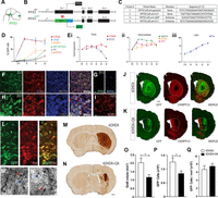
Loss of a striatal targets negatively impacts the size of hiPSC-derived VM progenitor grafts and the survival of DA neurons. Schematic sagittal view of the developing embryo illustrating the restrictive expression of PITX3, a key VM DA transcription factor (A). Monoalleleic targeting of enhanced GFP (eGFP) expression, driven under the PITX3 promotor region, used for the generation of a new PITX3-GFP iPSC cell line that enabled identification of VM DA neurons in vitro and in vivo (B). Specifically designed primers to amplify targeted PITX3 locus (C). Quantitative assessment of the proportion of cells expressing early (FOXA2, OTX2), intermediate (NURR1), and mature protein (TH, PITX3-GFP), reflective of VM progenitors and VM DA precursors and neurons (D). Quantitative PCR showing early (1), intermediate (2), and late (3) VM DA gene expression, confirming efficiency of the PITX3-GFP IPSC line to differentiate (E). Photomicrographs illustrating high expression of OTX2, FOXA2, and Nestin (F) as well as low proportions of off-target genes PITX2 and BARHL1 (G) confirm correct VM regional specification at day 13 of differentiation. By day 25, cultures show extensive GFP, TH, and FOXA2 co-expression (H) as well as expression of the DA precursor gene, NURR1 (I). Representative images show the placement of hiPSC-derived DA grafts (GFP+) within the 6OHDA (J) or 6OHDA+QA (K) lesioned striatum. After transplantation, the co-expression of TH with GFP confirms the robustness of the GFP+ reporter as a measure of VM DA neurons (L). GFP+ immunostaining reveals the impact of QA lesioning (N) on intrastriatal grafts of hiPSC-derived VM progenitors, compared with 6OHDA controls (M). High-magnification images from M, N illustrating A9-like (red arrow) and A10-like (blue arrow) DA neuron morphology within hiPSC-derived grafts (M', N'). Quantitative analysis reveals that ablation of striatal MSN results in significantly smaller grafts (O, p = 0.022), containing fewer GFP+ cells (P, p = 0.042) and consequently unchanged GFP+ cell density (Q, p = 0.494) in QA+6OHDA-lesioned compared with 6OHDA-lesioned animals. Data are mean ± SEM; *p < 0.05. n = 3 independent differentiations/time point (D, Ei–Eiii), n = 7–8/group (O–Q). Scale bars: 100 µm (F–I), 1 mm (J, K, M, N), and 50 µm (L, M', N').
For transplantation, D19 VM progenitors were injected into the striatum of mice that had received 6OHDA lesions of the midbrain (6OHDA; Fig. 4J) or 6OHDA midbrain lesions + QA-lesioning of the striatum (6OHDA+QA; Fig. 4K) three weeks prior. After six months, DARPP32 immunostaining again confirmed extensive loss of striatal MSNs in QA-lesioned animals (Fig. 4K) that was accompanied by visible atrophy of the nuclei (6OHDA: 12.73 ± 0.29 mm3, 6OHDA+QA: 5.52 ± 0.28 mm3; p < 0.0001), comparable to that previously observed for the rodent fetal tissue grafting studies shown in Figure 1C,H. Confirming validity of the new reporter stem cell line to identify DA neurons, >95% of the TH+ neurons within the grafts co-expressed GFP (Fig. 4L), enabling visualization of both the cell bodies within the graft core (Fig. 4M,N, delineated white lines) and more specifically patterns of graft-derived reinnervation of the host brain. Volumetric analysis of the graft core revealed significantly smaller graft sizes in the QA-lesioned mice (6OHDA: 2.02 ± 0.27; 6OHDA+QA: 1.16 ± 0.14 mm3; p = 0.022; Fig. 4O). Moreover, these grafts contained significantly fewer GFP+ DA neurons (6OHDA: 12 435 ± 1513; 6OHDA+QA: 8319 ± 902 GFP+ cells; p = 0.042; Fig. 4P), resulting in an unchanged density of GFP+ cells (6.66 ± 0.88 × 10³ vs 7.46 ± 0.66 × 10³ GFP+ cells/mm3; p = 0.494; Fig. 4Q).
In the absence of a striatal target hiPSC-derived dopaminergic grafts seek target connectivity beyond the striatum
Given the detrimental impact of MSN ablation on the size and survival of hiPSC-derived DA grafts, not observed in rodent fetal grafts, we assessed whether there was a concomitant effect on innervation. The PITX3-GFP hiPSC-derived dopaminergic cells within the grafts, transplanted into the 6OHDA-lesioned striatum, were capable of extensive re-innervation of the host striatum, spanning the rostral-caudal axis (Fig. 5A). In contrast, when the same GFP-expressing donor cells were placed into the QA-lesioned striatum their ability to re-innervate the striatum was greatly diminished (Fig. 5B), evidenced by a 44% decrease in innervation volume (6OHDA: 7.74 ± 0.82 mm3; 6OHDA+QA: 4.32 ± 0.51 mm3; p = 0.0045; Fig. 5C) and 51% decrease in the density of striatal innervation (6OHDA: 48.14 ± 2.51; 6OHDA+QA: 23.61 ± 1.91% GFP+ immunoreactive pixels; p < 0.0001; Fig. 5D). Where a strong correlation between GFP cell numbers and striatal innervation was observed for grafts placed into the intact striatum (6OHDA: R2 = 0.72, open circles), no similar correlation was observed when grafts were placed into the QA-lesioned striatum, despite the significant reduction in GFP+ cells (6OHDA+QA: R2 = 0.05, closed circles; Fig. 5E). In contrast, and in alignment with the rodent fetal grafting (Fig. 2), a strong inverse correlation between the extent of striatal lesioning (% DARPP32 lesion) and GFP innervation of the striatum (r2 = 0.74; Fig. 5F) was observed, with GFP+ neurons instead preferentially innervating the overlying cortex (Fig. 5Aiii,Aiv,Biii,Biv). Quantitative assessment revealed an 8.6-fold increase in the density of cortical innervation for grafts placed into QA-lesioned mice (6OHDA: 1.04 ± 0.25%; 6OHDA+QA: 8.97 ± 1.63% GFP+ immunoreactive pixels; Fig. 5G) and a correlative trend between the extent of the QA lesion (%DARPP32 lesion) and cortical innervation (r2 = 0.43; Fig. 5F).

hiPSC-derived dopaminergic neurons seek alternative connectivity in the absence of striatal targets. Representative photomontages illustrating the GFP+ fiber innervation patterns of hiPSC-derived DA neurons when grafted into the 6OHDA (A) and 6OHDA+QA (B) lesioned striatum. Darkfield imaging of GFP+ innervation of the forebrain (Ai, Bi, bregma: −0.5 mm), with high-magnification images of the striatum (Aii, Bii), somatosensory cortex (Aiii, Biii) and motor cortex (Aiv, Biv), illustrating the increased innervation of the cortex in 6OHDA+QA-lesioned animals. The ablation of MSN targets resulted in reduced striatal innervation volume (C, p = 0.0045) and density (D, p < 0.0001), supported by the lost correlation between the level of striatal innervation and the number of GFP+ cells observed in non-QA-lesioned animals (E, 6OHDA: r2 = 0.72 vs QA: r2 = 0.04). QA striatal lesioning (% DARPP32 lesion) inversely correlated with GFP striatal innervation (F, r2 = 0.74; black dots) and a converse correlation with cortical innervation (F, r2 = 0.43; red dots). G, QA lesioning resulted in an increase in cortical innervation by hiPSC-derived VM progenitor grafts. H, I, Representative photomontages illustrating the dense network of human synaptophysin (hSYP) synapses and GFP+ fibers within the striatum yet sparse labeling in the cortex of animals receiving grafts in the absence of a QA lesion. J, K, Conversely, grafts placed into the QA-lesioned animals displayed few GFP+ and hSYP+ synapses in the striatum, with notable increased cortical synaptic innervation. L, High-magnification image, from K, illustrating the colocalization of hSYP and GFP within the cortex of a QA-lesioned animal, indicating the formation of DA synapses. Striatum (STR); Cortex (CTX). Data are mean ± SEM; **p < 0.01, ***p < 0.001. n = 7–8/group. Scale bar: 1 mm (A, B).
Importantly, immunohistochemical labeling against synaptophysin protein, using a human-specific antibody (hSYP), confirmed that the GFP innervation resulted in mature synapses. In the absence of a QA lesion, dense networks of hSYP were observed throughout the striatum yet was notably absent within the overlying cortex (Fig. 5H,I). In comparison, hSYP immunoreactive puncta were rare in the striatum of animals receiving a QA lesioned but formed dense networks in the cortex (Fig. 5J,K), with evident co-localized GFP+hSYP+ fibers (Fig. 5L).
The loss of a striatal target significantly alters the dopaminergic phenotype acquisition of hiPSC-derived grafts
In view of the significant impact of striatal lesioning on target connectivity of the grafted hiPSC-derived neurons, we next sought to determine the effect on phenotype acquisition of the residing dopaminergic neurons. Reflective of our previous observations of human PSC-derived DA grafts (Niclis et al., 2017a; de Luzy et al., 2019, 2021; Gantner et al., 2020b), in control 6OHDA-lesioned animals the majority (56.26 ± 2.41%) of GFP+ cells co-expressed GIRK2, 4.86 ± 0.67% co-expressed Calbindin, and 35.13 ± 1.77% of the GFP+ cells failed express either protein, remaining GFP+/GIRK2–/CALB– (Fig. 6A,F). As previously observed for fetal and hPSC-derived DA grafts, DA neurons again retained a spatial organization with GIRK2+ cells at the graft periphery and CALB+ neurons at the core (Fig. 6A,A' and B,B', red and blue dots, respectively). While the regional distribution of GIRK2 and Calbindin-expressing DA neurons within grafts implanted into QA-lesioned animals was similar, striatal lesioning significantly reduced the proportion of GIRK2-expressing GFP+ cells (38.12 ± 2.53%), while conversely increasing the contribution of CALB+ cells (10.10 ± 1.20%; Fig. 6B,F). Added to the shift in A9 and A10-like proportions, a significant increase in the number of GFP+ cells failing to commit to a mature DA phenotype was observed in QA-lesioned animals (47.3 ± 3.3% GFP+/GIRK2–/CALB– cells; Fig. 6F), likely underpinned by the loss of the striatal MSN target, and capacity to adopt an A9-like GIRK2+ fate. More rigorous total A9/A10 cell counts highlighted a significant increase in the total number of Calbinin+GFP+ DA neurons within grafts implanted into the QA-lesioned striatum (908.7 ± 153.3), compared with grafts in the 6OHDA-lesioned animals (518.1 ± 58.79; Fig. 6G). This increase in total CALB+ cells strongly supports a fate shift of GFP+ cells, and we reason this may be a consequence of increased cortical target acquisition of developing neurons in light of lost striatal targets, and that the altered proportions (increase in %CALB+GFP+) do not merely reflect the loss of A9 DA neurons.

Loss of striatal target influences the acquisition of the A9 and A10 dopaminergic phenotype in hiPSC-derived dopaminergic neurons. Photomicrographs (A, B) and schematic reconstructions (A', B') illustrating the expression and distribution of DA phenotypes within grafts in the 6OHDA (A) or 6OHDA+QA-lesioned brain (B). Representative photomicrographs showing GFP+ cells co-expressing GIRK2 (Ci), CALB (Cii), and GIRK2/CALB (Ciii) within hiPSC-derived DA grafts. The absence of striatal MSNs resulted in a significant reduction in the proportion of A9-like GFP+/GIRK2+ and increase in A10-like GFP+/CALB+ DA neurons within hiPSC-derived VM grafts (p = 0.0008 and p = 0.0053, respectively; F, G), and an overall increase in GFP+ DA neurons that failed to mature into either phenotype (GFP+/GIRK2–/CALB–, p < 0.012; F, G). Representative images showing the high proportion of GIRK2+ cells co-expressing ALDH1A1, indicative of A9-like DA neurons of ventral tier of the substantia nigra pars compacta (D, E). Grafting into the QA-lesioned striatum significantly reduced the proportion of ALDH1A1+GIRK2+GFP+, but not ALDH1A1–GIRK2+GFP+ DA neurons (H). Reciprocally, a significant increase in A10-like neurons (ALDH1A1–CALB+GFP+) was observed (I). Data are mean ± SEM; *p < 0.05, **p < 0.001, ***p < 0.0001. n = 7–8/group (F, G); n = 3/group (H, I). Scale bars: 200 µm (A, B) and 100 µm (D, E).
The loss of A9-like DA neurons could be further validated by labeling for aldehyde dehydrogenase (ALDH1A1), a PITX3 target gene restricted to the ventral tier of the SNpc (the population that preferentially degenerate in PD (Pereira Luppi et al., 2021), and is necessary for restoration of motor function after grafting (Grealish et al., 2010). Unsurprisingly, ALDH1A1+ neurons within the graft showed a high degree of co-localization with GIRK2 (Fig. 6D,E), with few ALDH1A1+CALB+ cells present (reflective of a small population of neurons within the ventromedial region of the VTA; Fig. 6I). Similar to the loss of GIRK2+ neurons, a significant reduction in the proportion of ALD1A1+ was observed within grafts transplanted into the QA-lesioned striatum (6OHDA: 53.03 ± 7.14; 6OHDA+QA: 15.64 ± 2.12, p = 0.038; Fig. 6H). Reflective of their vulnerability in PD, these GIRK2+ALDH1A1+ DA cells, and not GIRK2+ALDH1A1– DA neurons cells were the population lost within the grafts.
Discussion
Neurodevelopmental studies have highlighted that a complex interplay of cell intrinsic and extrinsic cues are necessary for the establishment of neural circuitry in the developing brain. In the context of repair, neural grafts targeted at replacing dopamine neurons in the Parkinsonian brain are said to be “intrinsically programmed” to innervate the striatum (for review, see Herman and Abrous, 1994; Winkler et al., 2000; Thompson and Björklund, 2012). Here, we demonstrate, through selective ablation of the target MSNs of the striatum, the impact of extrinsic cues from the surrounding host environment that underpin survival, axonal remodeling, and fate specification of subpopulations of VM dopaminergic neurons.
During development VM DA neurons extend their axons toward their targets where they compete to form functional synapses. Availability of neurotrophic factors such as GDNF and BDNF at these targets means that only projecting neurons that make target contacts survive thus ensuring that the number of innervating neurons matches the size of the region in which they target, a phenomena known as the “Neurotrophic theory” (Barde, 1989). This is partly reflected in the context of neural transplantation where grafts placed into the aging brain show poor survival, compared with grafts into younger recipients, suggested to be a consequence of diminishing neurotrophic tone in the striatum (Collier et al., 1999; Sortwell, 2003). Conversely, survival is increased when grafts are placed into the striatum denervated of host DA terminals where trophic tone is elevated (Yurek and Fletcher-Turner, 2001) and there is increased availability of synaptic target sites present (Doucet et al., 1990; Kirik et al., 2001). In the present findings we show that ablation of the target striatal MSNs had no influence on the size or DA neuron number for grafts derived from rodent fetal tissue, reflective of previous observations (Duan et al., 1998), yet had a significant impact on hiPSC-derived VM progenitor grafts. We propose that the discrepancy reflects differences in total DA neuron numbers across the two grafting cohorts, whereby fewer DA neurons in fetal tissue grafts compete for residual target MSN neurons, compared with the 10-fold greater number of surviving DA neurons in hiPSC-derived grafts. Future studies, involving a more detailed dose response of DA neurons (from rodent or human and/or fetal or PSCs) as well as varying QA striatal lesion sizes will be required to verify and assess the critical threshold for graft to host target ratios underlying cell survival. Added to this is the consideration as to whether the differences in survival may also reflect the origin of the donor cells (fetal vs pluripotent stem cells) and/or xenogeneic grafting influences.
To what extent cell intrinsic versus extrinsic cues drive synaptic connectivity remains unclear. Are cells preprogrammed to target a specific cell type, and/or are specific cues, inclusive of secreted and cell adhesive proteins from the surrounding environment responsible for target acquisition? The present findings showing DA fibers re-routing from the striatum to the cortex following ablation of the striatal MSNs highlights the significant role of extrinsic cues. The results complement previous studies in which both transgenic models and the delivery of developmentally appropriate proteins into the target tissues emphasize the importance of cell extrinsic signals, for example, the overexpression of GDNF (Gantner et al., 2020b) and conversely loss of cell adhesive proteins such as ALCAM (Bye et al., 2019) increase and decrease, respectively, the plasticity of DA neurons within grafts. Extrinsic cues from the cortex, however, are not in themselves sufficient to attract more graft-derived DA fibers (or influence survival of hiPSC-derived grafts). One is also left to speculate whether the intrinsic drive from the DA neurons within the grafts also limits the extent of the alternative mesocortical trajectory.
Reflective of the altered innervation levels and synaptic targeting by the grafted neurons implanted into the 6OHDA-lesioned or 6OHDA+QA-lesioned striatum was a shift in DA fate specification. Within the adult VM, the A9 subpopulation of DA neurons is critical for motor function, with the degeneration of these neurons underpinning the motor symptoms in PD (for review, see Björklund and Dunnett, 2007). In correlation, the presence of these A9-like neurons are critical for VM grafts to restore motor function (Kuan et al., 2007), with their selective removal from grafts resulting in a failure to reverse motor impairments (Grealish et al., 2010). While a series of studies have identified that the A9 DA neurons precede the birth of A10 (Blaess et al., 2011; Hayes et al., 2011; Bye et al., 2012), and consequently transplantation of younger fetal tissue can selectively bias this population within grafts (Bye et al., 2012), no distinguishing gene/s or protein/s have been able to discriminate these subpopulations during their development, or enable targeted generation from PSC sources in vitro. Here, we describe evidence for a model where target acquisition plays a role in A9 and A10 fate specification. In the absence of striatal targets, both fetal and hiPSC-derived DA neurons showed a significant reduction in A9-like GFP+GIRK2+ DA neurons and a commensurate increase in the proportion of DA neurons that failed to adopt either an A9-like or A10-like fate. Furthermore, the 8.5-fold increase in cortical innervation was commensurate with a significant increase in A10-like Calbindin-expressing neurons for hiPSC-derived grafts for which significantly more DA neurons were present and void of targets than fetal grafts. Future studies, involving retrograde tracing from the dorsolateral striatum or cortex, will be important to validate a direct correlation between re-routed axonal fibers and the impact on DA subtype specification. It should also be considered that the inflammatory environment following QA lesioning may impact fate specification, although it seems unlikely this would selectively impact the A9 population while leading to an increase in A10 maturation. Nonetheless, future studies involving the delivery of compounds that induce focal inflammatory responses (such as lipopolysaccharide), without ablation of the striatal target neurons, should be undertaken to address this possibility.
In summary, we present a series of findings that highlight the importance of target acquisition for the survival, plasticity, selective targeting, and fate specification of VM DA neurons. We demonstrate these largely maintained effects in both allogeneic grafts (fetal mouse grafts into mice) as well as xenogeneic grafts (human into mouse) suggesting a conserved mechanism across species, and the assumption that similar target acquisitions are necessary in the establishment of the developing human midbrain networks and clinical trials involving human donor tissue, whether it be of fetal or hPSC origin.
Footnotes
- Received December 12, 2021.
- Revision received April 14, 2022.
- Accepted April 15, 2022.
-
This work was supported by the National Health and Medical Research Council (NHMRC) Australia. C.P. was supported by an NHMRC Australia Senior Research Fellowship. I.R.d.L. and V.P. were supported by University of Melbourne International Scholarships, Australia. The Florey Institute of Neuroscience and Mental Health acknowledges the strong support from the Victorian Government and in particular the funding from the Operational Infrastructure Support Grant. We thank Ms. Brianna Xuereb and Ms. Mong Tien for their expert technical assistance.
-
The authors declare no competing financial interests.
- Correspondence should be addressed to Clare L. Parish at clare.parish{at}florey.edu.au or Lachlan H. Thompson at lachlan.thompson{at}florey.edu.au
- Copyright © 2022 the authors




