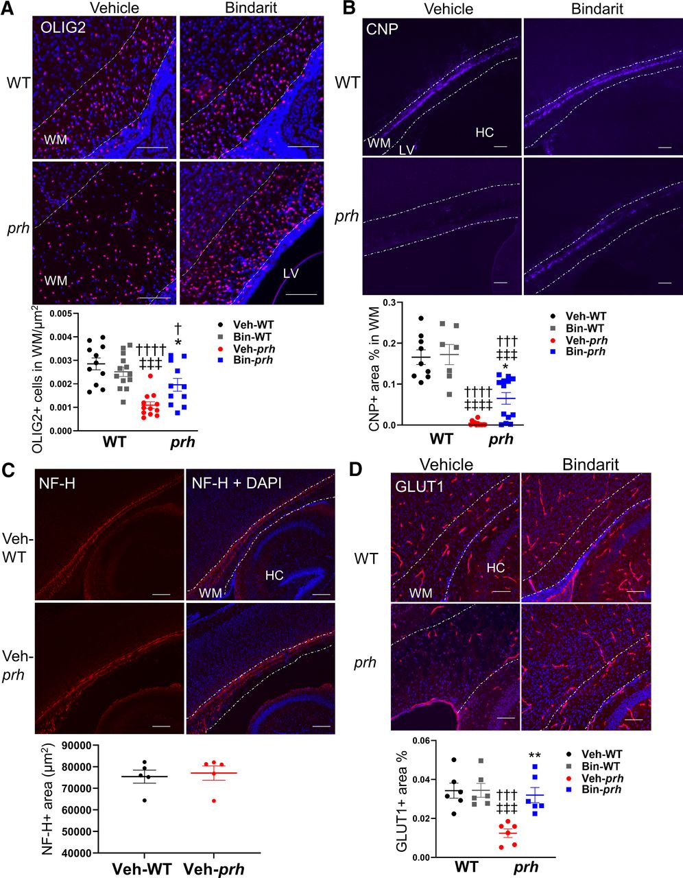
Myelination and vascularization in prh white matter with bindarit treatment. A, Number of pan-oligodendrocyte lineage marker OLIG2+ cells per WM shows reduced oligodendrocyte density in veh-prh and improvement in bin-prh (†p = 0.0367, ††††p < 0.0001 vs veh-WT, ‡‡‡p = 0.001 vs bin-WT, *p = 0.0385 vs veh-prh; two-way ANOVA followed by Tukey's test, n = 12–13 in each group; scale bars: 100 μm). B, The mature myelination marker CNP positive staining area % in WM shows significant reduction of myelination in veh-prh and partial recovery in bin-prh (†††p = 0.0001, ††††p < 0.0001 vs veh-WT, ‡‡‡p = 0.0002, ‡‡‡‡p < 0.0001 vs bin-WT, *p = 0.0133 vs veh-prh; two-way ANOVA followed by Tukey's test, n = 10–13 in each group; scale bars: 100 μm). C, Neurofilament-labeled axons (NF-H) positive area (μm2) in WM shows no significant difference between veh-WT and veh-prh (p = 0.7244; Student's t test, n = 5 in each group; scale bars: 200 μm). D, Endothelial cell marker, GLUT1 positive staining area per WM (%) shows reduced vascular densities in veh-prh and significant recovery in bin-prh (†††p = 0.0004 vs veh-WT, ‡‡‡p = 0.0001 vs bin-WT, **p = 0.0019 vs veh-prh; two-way ANOVA followed by Tukey's test, n = 6 in each group; scale bars: 100 μm). WM, white matter; LV, lateral ventricles; HC, hippocampus.


