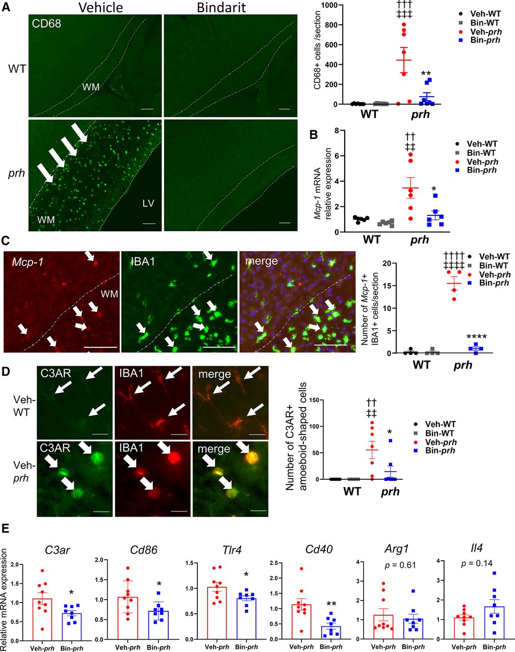
Bindarit's effect on myeloid cell activation in the periventricular white matter. A, CD68+-activated myeloid cells are rarely seen in veh-WT and bin-WT at P8, but significantly increased in white matter (WM) of veh-prh, which is significantly suppressed in bin-prh (†††p < 0.0001 vs veh-WT, ‡‡‡p < 0.0001 vs bin-WT, **p = 0.0040 vs veh-prh; two-way ANOVA followed by Tukey's test, n = 6–7 in each group; scale bars: 100 μm). B, Mcp-1 mRNA relative to Gapdh mRNA is upregulated to 3.47 ± 0.82 times relative to veh-WT in veh-prh and is significantly suppressed in bin-prh (†††p = 0.0047 vs veh-WT, ‡‡‡p = 0.0016 vs bin-WT, **p = 0.0135 vs veh-prh; two-way ANOVA followed by Tukey's test, n = 6 in each group; scale bars: 100 μm). C, Mcp-1 in situ hybridization shows exclusive Mcp-1 mRNA expression by amoeboid-shaped IBA1+ myeloid cells in the periventricular WM and lower cortical layers of veh-prh, and significant suppression in bin-prh (††††p < 0.0001 vs veh-WT, ‡‡‡‡p < 0.0001 vs bin-WT, ****p < 0.001 vs veh-prh; two-way ANOVA followed by Tukey's test, n = 4 in each group; scale bars: 100 μm). D, Complement receptor C3AR is weakly expressed in IBA1+ ramified microglia in gray matter of veh-WT (top) but is strongly expressed in amoeboid-shaped IBA1+ cells in WM of veh-prh (bottom). Number of C3AR-expressing amoeboid-shaped IBA1+ cells is suppressed in bin-prh relative to veh-prh (††p = 0.0027 vs veh-WT, ‡‡p = 0.0027 vs bin-WT, *p = 0.0325 vs veh-prh; two-way ANOVA followed by Tukey's test, n = 7 in each group; scale bar: 50 μm). E, Proinflammatory genes and cytokines involved in microglia/macrophage activation; C3ar (*p = 0.0437), Cd86 (*p = 0.0468), Tlr4 (**p = 0.0052), and Cd40 (*p = 0.0456) mRNA are significantly suppressed in bin-prh compared with veh-prh, while alternative activation (M2) markers of microglia/macrophages Arg1 (p = 0.61) and Il4 (p = 0.14) are not changed by bindarit (Student's t test, n = 8–9 in each group).


