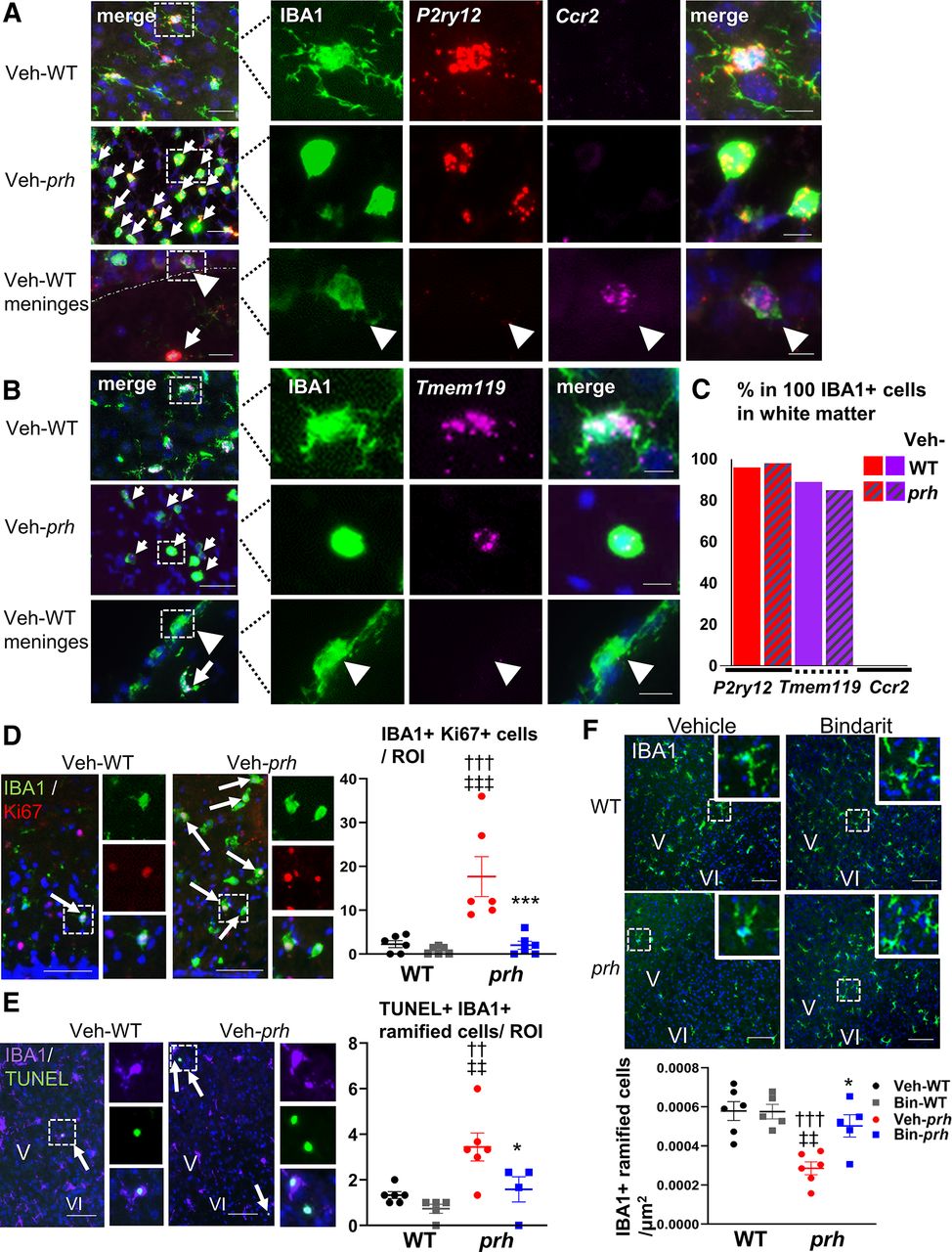
Bindarit suppresses microglial activation in the white matter and restores the ramified microglia in the prh gray matter. A, The microglial signature gene P2ry12 (purinergic receptor P2Y, G-protein-coupled 12) is expressed on ramified-shaped IBA1+ cells in WTs at P8 (top row). The amoeboid-shaped IBA1+ cells in the white matter (WM) of veh-prh at P8 also express P2ry12 to a lesser extent, but do not express the monocyte signature gene Ccr2 [chemokine (C-C motif) receptor 2; middle row]. The Ccr2 probe is validated, and its signal is observed on IBA1+ cells outside the brain in veh-WT at P8, which do not have P2ry12 signal (bottom row, arrowhead), whereas P2ry12 is expressed on cortical microglia on the same slide (bottom row, arrow; n = 3–4; scale bars: 50 μm for low-magnification pictures, 10 μm for high-magnification pictures). B, Another microglial signature gene Tmem119 is expressed on ramified-shaped IBA1+ cells in veh-WT at P8 (top row). The amoeboid-shaped IBA1+ cells in the WM of veh-prh at P8 also express Tmem119 to a lesser extent (middle row). Tmem119 is not expressed on IBA1+ cells outside the brain in veh-WT at P8 (bottom row, arrowhead), whereas cortical microglia express Tmem119 on the same slide (bottom row, arrow; n = 2; scale bars: 50 μm for low-magnification pictures, 10 μm for high-magnification pictures). C, Graph shows the quantification of P2ry12 (96%, 98%), Tmem119 (89%, 85%), and Ccr2 (0%, 0%) expression in 100 IBA1+ cells (n = 2–3) in the WM of veh-WT and veh-prh, respectively. All 100 IBA1+ cells quantified in veh-prh are amoeboid-shaped cells. D, Double-staining of IBA1 and the proliferation marker Ki67 shows very few double labeled cells in the WM of veh-WT (top) but significantly more proliferating amoeboid-shaped IBA1+ cells (arrows) in the WM of veh-prh (bottom; †††p = 0.0009 vs veh-WT, ‡‡‡p = 0.0003 vs bin-WT). This effect is suppressed in bin-prh brains (***p = 0.0008, two-way ANOVA followed by Tukey's test, n = 6 in each group; scale bars: 100 μm). E, TUNEL assay with IBA1 staining shows significantly more TUNEL positive (i.e., dying) ramified microglia (arrows) within the gray matter of veh-prh cortex (††p = 0.0082 vs veh-WT, ‡‡p = 0.0014 vs bin-WT). Bindarit treatment significantly reduces this cell death in bin-prh cortex (*p = 0.0411 vs veh-prh; two-way ANOVA followed by Tukey's test, n = 4–6 in each group; scale bars: 100 μm). F, The density of ramified microglia within the gray matter (μm2) is significantly reduced in veh-prh (†††p = 0.0006 vs veh-WT, ‡‡p = 0.0012 vs bin-WT), and this reduction is significantly restored in bin-prh cortex (*p = 0.0140 vs veh-prh; two-way ANOVA followed by Tukey's test, n = 6 in each group; scale bars: 100 μm).


