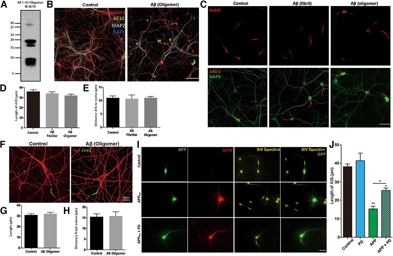
APP-induced changes in the AIS are independent of Aβ but partially dependent on calpain activity. A, Western blot of the Aβ oligomer sample used (blotted with the 6E10 antibody). B, DIV14 primary cultured neurons treated with Aβ oligomer (1 μm) for 24 h, then fixed and immunolabeled for Synapsin1 (red), Aβ (6E10 antibody, green), and MAP2 (cyan). Nuclei were labeled with DAPI (blue). Scale bar, 20 µm. C, DIV14 neurons treated with fibrillar or oligomeric Aβ for 24 h, then immunolabeled for AnkG (red) to reveal the AIS and for MAP2 (green). Scale bar, 25 µm. D, Quantification of the average length of the AIS in cultures, such as those in C. E, Quantification of the distance of the AIS from the soma. F, Glutamatergic neurons were generated from human iPS cells. After 4 weeks maturation, the cells were treated with Aβ oligomers for 24 h before fixation and immunostaining with Tuj1 (red) and AnkG (green). Scale bar, 10 µm. G, Quantification of the AIS length in cultures, such as that shown in F. H, Quantification of the distance of the AIS from the soma. I, DIV7 neurons transfected with APPSwe plasmid with or without the calpain inhibitor, PD150606 (PD), for 2 d and immunolabeled with the AIS marker βIV spectrin (yellow) and APP (6E10, red). GFP labels the transfected cells and is shown in green. Scale bar, 20 µm. J, Quantification of the length of the AIS from the experiment shown in I. Symbols over individual bars represent significance of difference from control. Data are mean ± SEM. *p < 0.05; **p < 0.001; one-way ANOVA. n = 4 batches of independent cultures.


