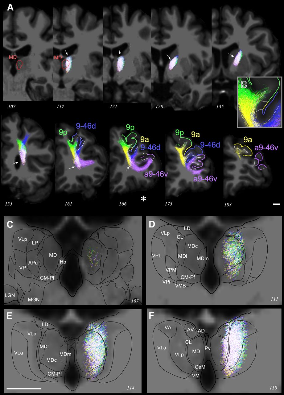
Intrathalamic and extrathalamic paths of the tracts connecting MD with four cortical areas belonging to the same dPFC group region. A, Streamline visualizations of the tractographies between MD (red line contour) and the inferior transitional area between areas BA9 anterior (9a; yellow) and BA9 posterior (9p; green); the transitional area between BA9 and dorsal BA46 (9-46d; blue); the anterior transitional area between BA9 and ventral BA46 (a9-46v; purple). The part of the cortex corresponding to each seed area is delineated, and its pial surface is highlighted in color. Each tract is coded in the corresponding color; white indicates coregistration of all four streamlines in the same voxel. Note that the four tracts remain strictly overlapped (arrow) from their origin in MD and in the internal capsule, all the way into the pallial white matter. There, the four tracts split abruptly to follow independent trajectories toward their target areas. The asterisk indicates the coronal slice from which the magnified image shown in B was taken. B, Magnified visualization of a coronal slice showing the streamlines reaching cortical areas 9p, 9a, and 9-46d. C–F, Coronal sections of both thalami at higher magnification. Tractographies were obtained from the same individual shown in Figure 2. Note that each of the four tracts remain coregistered extensively in the same voxels as they traverse the gray matter of the thalamus and enter MD. Scale bars: A, 1 cm; E (for B–F), 1 cm. Graphic conventions and abbreviations are as in Figure 3.


