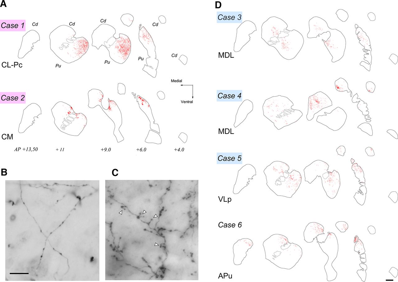
Thalamostriatal projections from CT nuclei and summary diagram. A, Section diagrams of the caudate (Cd) and putamen (Pu) taken at five representative levels of the striatum are shown to illustrate the thalamostriatal axon arborizations (red stippling) labeled by injections in the anterior/dorsal (Case 1) or posterior/ventral (Case 2) portions of the IML. The levels rostral to the interaural coronal plane (AP+) are indicated. B, High-magnification image of thalamostriatal axon arborizations labeled by the CL–Pc injection. Note the diffuse and linear arrangement of the axonal branches and the presence of en passant boutons in them. C, Thalamostriatal arborizations labeled by the BDA injection in CM–Pc. Note the focal, plexus-like morphology of the terminal branches and frequent terminal boutons at the end of a short stalk. D, Thalamostriatal axon arborizations labeled from injections in regions of CT with sparse projections to the striatum. The labeling observed by the injection in Case 6 (APul) is also shown. Conventions are as in A. Scale bars: A–D, 1 mm; B, C, 10 µm.


