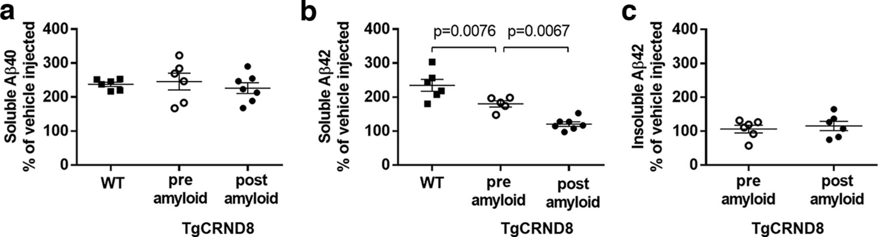
Sensitivity of synaptosomal Aβ40 and Aβ42 to degradation by ECE-related proteases in TgCRND8 mice, pre- and post-amyloid deposition. Mice were injected intracerebroventricularly with phosphoramidon (PA) or saline vehicle. Crude synaptosomes were isolated 16 h later and extracted sequentially with RIPA and GuHCl for measurement of Aβ by sandwich ELISA. a, RIPA-soluble synaptosomal Aβ40 was elevated similarly in WT mice (WT, n = 6, 5 M/1 F) and in TgCRND8 mice pre-amyloid deposition (n = 6, 2 M/4 F, aged 8-10 weeks) or post-amyloid deposition (n = 7, 3 M/4 F, aged 16-17 weeks) (F(2,16) = 0.33, p = 0.72, ANOVA). b, There was significantly less PA-induced accumulation of RIPA-soluble synaptosomal Aβ42 in young TgCRND8 mice compared with WT mice, and a further reduction in accumulation in TgCRND8 mice after plaque deposition (F(2,15) = 24.75, p < 0.0001, ANOVA). Multiplicity-adjusted p values (Holm–Sidak's) are indicated on the graph. c, PA treatment did not significantly affect RIPA-insoluble (GuHCl-solubilized) Aβ42 in either age group. Data are plotted for each PA-treated group, expressed as a percent of the mean of age-matched vehicle controls which included WT (n = 6, 4 M/2 F), TgCRND8 pre-deposition (n = 6, 2 M/4 F), and TgCRND8 post-deposition (n = 7, 3 M/4 F). Lines indicate mean ± SEM.


