Erratum: Zhong et al., “Protein S Protects Neurons from Excitotoxic Injury by Activating the TAM Receptor Tyro3–Phosphatidylinositol 3-Kinase–Akt Pathway through Its Sex Hormone-Binding Globulin-Like Region”
In the article “Protein S Protects Neurons from Excitotoxic Injury by Activating the TAM Receptor Tyro3–Phosphatidylinositol 3-Kinase–Akt Pathway through Its Sex Hormone-Binding Globulin-Like Region,” by Zhihui Zhong, Yaoming Wang, Huang Guo, Abhay Sagare, José A. Fernández, Robert D. Bell, Theresa M. Barrett, John H. Griffin, Robert S. Freeman, and Berislav V. Zlokovic, which appeared on pages 15521–15534 of the November 17, 2010 issue, there is an error in Figure 2C. The authors note the following:
“We determined that there were similarities between the Akt loading control blots in Fig. 2C and the β-actin loading control blots in Fig. 5E, which was due to a mistake in the selection of the Akt control blots for Fig. 2C during manuscript preparation. Since we do not have the original Western blots used in Fig. 2C from approximately 15 years ago, we repeated the experiment shown in Fig. 2C and obtained similar results. Although the error was only in the original Akt control blot, all three blots shown in Fig. 2C were repeated so that the quantification of pGSK3α/β abundance could be correctly normalized to the matching new Akt control blots, following the methods described in the original paper. The corrected Fig. 2C is shown below. For clarity, we also present 3 full blots generated and used for quantification when we repeated the experiment. We sincerely apologize for this unintentional error.”
This correction does not affect the conclusions of the paper. A corrected version of the figure is displayed below, as well as a new figure with full blots to help provide context.
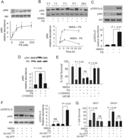
PS protects NMDA-treated mouse neurons via activation of PI3K/Akt antiapoptotic pathway. A, PS dose-dependently (5–200 nm) mediates Akt phosphorylation (pAkt, Ser473) in mouse cortical neurons 1 h after NMDA. Graph, pAkt relative abundance normalized to total Akt. *p < 0.05, PS compared with untreated NMDA controls. B, Akt phosphorylation (pAkt, Ser473) in mouse cortical neurons 1–24 h after NMDA with and without PS. Graph, pAkt signal normalized by total Akt. C, Akt activity determined by a GSK-3α/β phosphorylation-based assay 1 h after NMDA in the absence and presence of PS. pAkt and total Akt levels were determined by immunoblotting analysis from the same samples used in Akt activity assay. Graph, Relative abundance of pGSK-3α/β normalized by total Akt. D, Akt phosphorylation (pAkt, Ser 473) and total Akt levels in mouse cortical neurons 24 h after NMDA with or without PS or the PI3K inhibitor LY294002 (50 μm). E, Survival of mouse cortical neurons 24 h after NMDA with and without PS in the presence and absence of LY294002 (50 μm) or U0126 (10 μm). LY294002 and U0126 were added to the culture 30 min before NMDA treatment. F, Akt kinase activity in neurons transduced with kinase-deficient Ad.AktK179A mutant or control Ad.GFP determined 1 h after NMDA with and without PS by using a GSK-3α/β phosphorylation-based Akt activity assay. Graph, Relative abundance of pGSK-3α/β normalized by total Akt. pAkt and total Akt levels are also shown. G, Survival of DIV7 and DIV21 mouse cortical neurons transduced with Ad.AktK179A mutant or control Ad.GFP 24 h after NMDA with and without PS. Values are mean ± SEM from three to six independent cultures. In all studies, DIV7 cortical neurons and murine PS at 100 nm were used unless otherwise specified.

For clarity, the authors have also provided three full blots generated and used for quantification in Figure 2C when they repeated the experiment. Dashed boxes indicate the blots used for representative images in the corrected Figure 2C.
- Copyright © 2024 the authors




