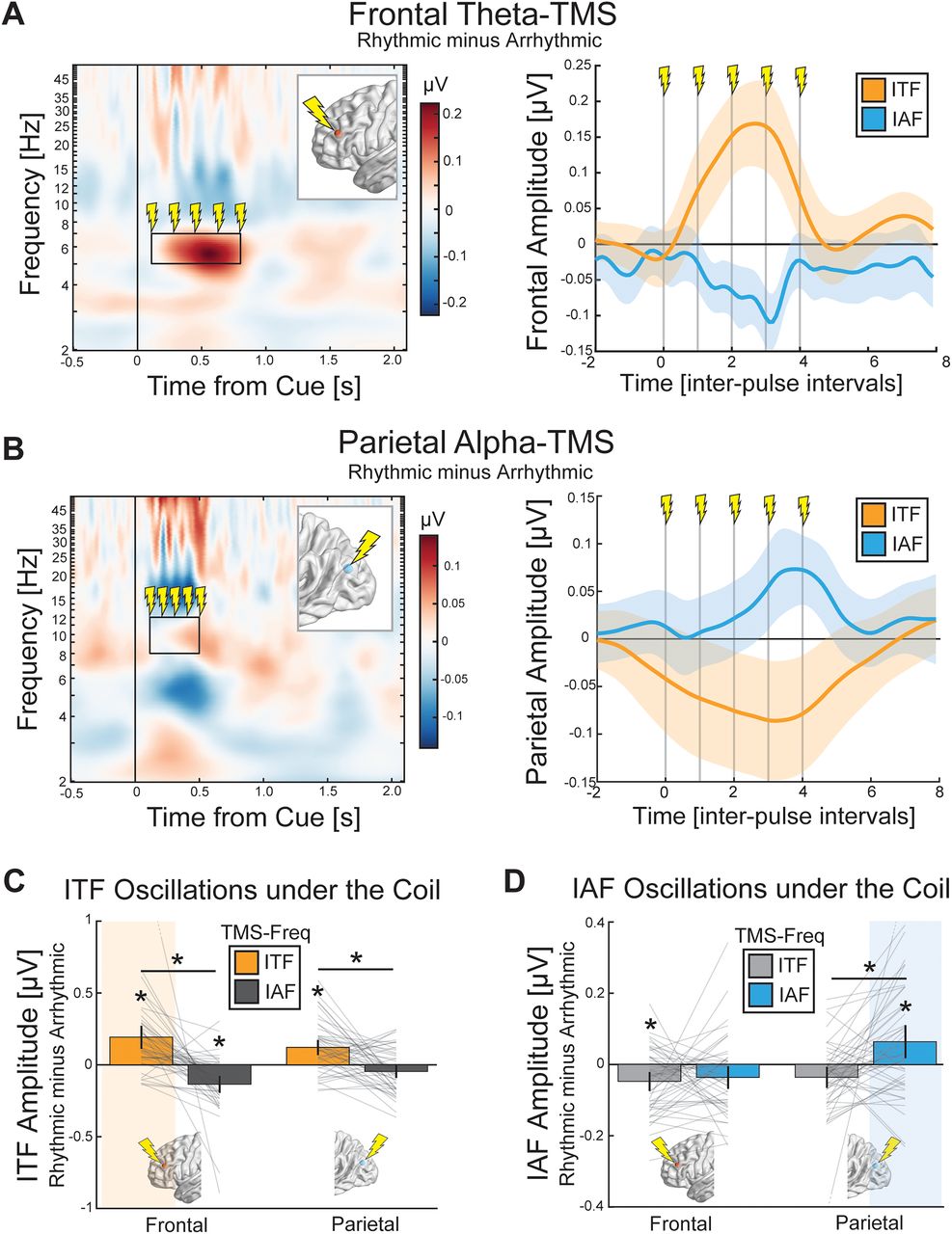
Rhythmic TMS increased the amplitude of the targeted neural oscillation. A, Theta-TMS to the frontal cortex delivered after cue onset (vertical black line) increased theta amplitude relative to arrhythmic TMS matched for duration and number of pulses (left). The targeted region is depicted in the inset. The black rectangle highlights the time and frequency range of delivered rhythmic TMS. Lightning bolts depict the approximate time of each TMS pulse. The amplitude of ITF and IAF during frontal theta-TMS was estimated, and amplitude modulation peaked at the fourth TMS pulse (right). B, Alpha-TMS to the parietal cortex increased alpha amplitude (left) with peak effects around the fifth TMS pulse (right). The vertical gray lines depict each TMS pulse in the burst. The shaded area is a 95% confidence interval relative to arrhythmic TMS. The amplitude estimation for the panels on the right were corrected for the individualized frequency of stimulation. C, The amplitude of ITF oscillations in the stimulated region as a function of the TMS site and TMS frequency revealed a robust increase in ITF amplitude when targeting both the frontal and parietal cortexes. D, IAF amplitude in the targeted region was increased from alpha-TMS in the parietal cortex, but not the frontal cortex. Error bars are SEM. *p < 0.05 after Bonferroni’s correction. The highlighted area depicts the condition hypothesized to show maximal target engagement.


