Vesicle Fusion in Oligodendrocyte Maturation and Myelination
-
Review of Fekete et al.
Introduction
The generation of myelin in the central nervous system requires the coordination of two poorly understood cellular processes: (1) cellular differentiation of oligodendrocytes from progenitors and (2) generation and trafficking of the lipids required to create myelin sheaths. As oligodendrocyte progenitor cells (OPCs) differentiate, they first enter a transitory “premyelinating” state in which cytoplasmic processes arborize extensively and contact potential targets for wrapping. A fraction of these contacts stabilize around axons and begin to concentrically envelop the axon through extensive membrane expansion and compaction. This results in the mature oligodendrocyte morphology (Fig. 1).
Previous studies have suggested that maturation from premyelinating to myelinating oligodendrocytes is a demanding cellular transition. Notably, cells at this stage begin generating new membranes in large quantities leading to high levels of cellular stress and up to half of premyelinating oligodendrocytes undergoing apoptosis (Barres et al., 1992). How do the surviving oligodendrocytes generate and traffic such large amounts of membrane? Another remarkable feature of the oligodendrocyte lineage is that OPC density is maintained by cellular division when neighboring cells differentiate or undergo apoptosis (Hughes et al., 2013). This may depend on autocrine signaling between cells at different stages of development (Fekete and Nishiyama, 2022), but this remains relatively unexplored. Do premyelinating cells communicate with nearby OPCs to regulate differentiation?
A trio of recent papers suggest the answers to the two questions posed above are related. In a study published in the Journal of Neuroscience, Fekete et al. (2023) found that vesicle trafficking and fusion may link OPC differentiation and lipid membrane expansion, particularly at the vulnerable stage of premyelinating oligodendrocyte maturation. Two other recent studies on the role of exocytosis in oligodendrocyte differentiation (Lam et al., 2022; Pan et al., 2023) provide a complementary set of mechanisms to explain how vesicle and membrane trafficking may contribute to myelination.
Myelin sheaths are composed of approximately 70–85% lipids and 15–30% proteins (Nornton and Cammer, 1984), but how these components are delivered to the correct target and form a sheath remains unclear. Previous work has suggested that the trafficking of cargo necessary for myelin production is regulated by SNARE proteins, a class of proteins required for the trafficking and fusion of vesicles in all eukaryotic cells (Feldman et al., 2011). To address how SNARE proteins and vesicular trafficking regulate oligodendrocyte development, Fekete et al. used a transgenic mouse line known as “iBot” to express botulinum neurotoxin light chain (BoNT/B-LC) in OPCs. This construct cleaves VAMP1-3 (a vesicular component of the SNARE complex) and thus prevents intracellular SNARE-mediated processes including vesicle fusion and targeting. The authors then asked how blocking VAMP2/3-dependent vesicle functions in OPCs affects oligodendrocyte lineage cells during development.
Compared with control mice, iBottg/+ mice expressing BoNT/B-LC in OPCs had a smaller population of mature oligodendrocytes identified by QKI7, a selective label for these cells (Bin et al., 2016). However, BoNT/B-LC expression did not affect the number of PDGFRɑ+ OPCs; this suggests that the survival and maturation of oligodendrocytes was impaired by the cleavage of VAMP2/3. In line with the decreased density of oligodendrocytes, mutant (iBottg/+) mice exhibited severe hypomyelination in the spinal cord—as assessed by immunofluorescence, Western blot, and qRT-PCR—and showed significant motor deficits prior to premature death.
To determine when during the process of differentiation oligodendrocytes are lost, Fekete et al. used EdU pulse-chase labeling to fate-map OPCs that were actively undergoing cell division at postnatal day 10 (P10). The population of OPCs labeled in iBottg/+ mice successfully differentiated into early, premyelinating oligodendrocytes by P13 but produced a significantly lower number of mature oligodendrocytes compared with that in the control. Thus, VAMP2/3 cleavage appeared to inhibit the maturation of newly formed oligodendrocytes rather than prevent the initial differentiation of OPCs. To further delineate the timing of oligodendrocyte loss, the authors examined the dynamic expression of the transcription factor NKX2.2, which shows a parabolic expression pattern across the oligodendrocyte lineage: it is upregulated in OPCs right before differentiation, immediately downregulated at the end of differentiation, and then re-expressed at low levels in mature oligodendrocytes. Using NKX2.2 as a readout for cellular maturity, Fekete et al. found that the number of NKX2.2+ mature oligodendrocytes was reduced in iBottg/+ mice, while the number of actively differentiating oligodendrocytes did not change. This further suggests that the maturation of premyelinating to mature oligodendrocytes is most affected when VAMP2/3 is cleaved. Finally, the authors demonstrated the iBottg/+ oligodendrocytes that fail to mature ultimately undergo cell death by apoptosis.
The study by Fekete et al. characterizes the diminished capacity of oligodendrocytes to reach a fully mature, myelinating state in the absence of functional VAMP2/3 protein. However, the multistaged nature of the oligodendrocyte lineage means that in vivo approaches are limited by the cell-state–specific promoters used to drive transgenes (Fig. 1) such that parallel strategies are often required to paint a complete picture of the biology at hand. Each Cre line drives transgene expression at slightly different times in oligodendrocyte development and with different degrees of cellular specificity, both of which should be considered when interpreting experimental results. Fekete et al. used the NG2-Cre mouse line, which targets all OPCs as well as pericytes, a cell type that is tightly associated with the vasculature.
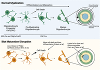
Top, Oligodendrocyte precursor cells (OPCs), targeted by NG2-Cre and Pdgfra-CreER, differentiate to form premyelinating oligodendrocytes and then mature into myelinating oligodendrocytes, with later stages labeled by CNP-Cre. During the maturation of these oligodendrocyte lineage cells, large amounts of lipid membrane must be deposited to expand the myelin sheath longitudinally by trafficking vesicles (black dots) concentrically around the ensheathed axon. Bottom, Fekete et al. and others found that in “iBot” mice with disrupted Vamp2/3-dependent vesicle fusion, premyelinating oligodendrocytes undergo cell death at higher rates rather than forming mature oligodendrocytes. Proposed mechanisms for the deficit include impaired vesicular release of signaling factors to regulate differentiation (left side) and decreased fusion of lipid-bearing vesicles to grow the expanding myelin sheath (right side). Figure generated using biorender.com.
Beyond this caveat, the results of Fekete et al. do not explain the molecular mechanism underlying defects in oligodendrocyte maturation. In the mammalian brain, VAMP2/3 is best known for its role in synaptic vesicle docking during exocytosis. If VAMP2/3 functions in a similar manner in oligodendrocyte lineage cells, it is reasonable to predict that targeting VAMP2/3 in OPCs will limit exocytosis and thus inhibit any autocrine signaling that may promote differentiation and maturation. Alternatively, inhibiting vesicular fusion may prevent the targeted delivery of lipids to the exterior membrane of oligodendrocytes during the active phase of myelin formation. Fortunately, two other recent studies using genetic approaches similar to those of Fekete et al. provide further mechanistic insight.
The potential role of autocrine signaling factors in the maturation of oligodendrocytes was investigated by Pan et al. (2023). Like Fekete et al., Pan et al. used the iBottg/+ mouse line to disrupt SNARE-mediated fusion in OPCs, but they targeted OPCs using the Pdgfra-CreER mouse line, which allows tamoxifen-inducible expression in OPCs and perivascular fibroblasts but not in pericytes. These mice exhibited strong myelination deficits in the brain during development, corroborating results shown by Fekete et al. Pan et al. then modeled this result in vitro, where iBottg/+ OPCs failed to differentiate or expand their membranes normally. Remarkably, however, when media from control oligodendrocyte lineage cells was provided to iBottg/+ OPCs, deficits in maturation were ameliorated suggesting a secreted factor signals in an autocrine manner to promote the maturation of newly formed oligodendrocytes. To identify a specific secreted factor that may be at play, Pan et al. mined a list of genes encoding secreted molecules that are highly expressed in oligodendrocytes. They selected Ptgds, a gene that encodes an enzyme involved in prostaglandin synthesis, for further investigation. Pan et al. then used both culture supplementation and genetic overexpression to show that the prostaglandin pathway regulates oligodendrocyte maturation. Thus, part of the mechanism through which VAMP2/3 cleavage impairs myelination appears to be through inhibiting the release of an autocrine signaling factor that normally encourages oligodendrocyte maturation.
To investigate the role of SNARE proteins in lipid trafficking, Lam et al. (2022) crossed the iBottg/+ mouse line with a Cnp-Cre line. This Cre line targets the oligodendrocyte lineage from the stage when OPCs begin to differentiate through maturity and is also known to target spinal motor neurons. Using this line, they tested whether oligodendrocyte maturation—independent of effects on the earlier OPC stage—requires VAMP2/3-mediated lipid addition. First, they asked whether VAMP2/3-associated vesicles are involved in lipid delivery at the myelin membrane. Imaging of a pH-sensitive fluorescent tag conjugated to SNARE proteins in cultured Cnp-iBottg/+ oligodendrocytes revealed that most exocytosis events resulted in a full fusion of the vesicular membrane with the cell surface, as opposed to brief fusion with the membrane and reformation of the vesicle, with full fusion associated with release of vesicular content. This suggests that vesicular lipids are efficiently transferred to the oligodendrocyte cell membrane and thus may contribute to myelin generation. Furthermore, in vivo imaging in larval zebrafish showed that VAMP2/3 exocytosis events were particularly prevalent during the premyelinating stage and were disproportionately targeted to the edge of the myelin sheath, where there is likely great demand for lipid incorporation. Electron microscopy revealed an accumulation of vesicles at the inner tongue of myelin sheaths in Cnp- iBottg/+ mice, suggesting that these vesicles failed to fuse in the growing sheath in the absence of VAMP2/3. Collectively, these results suggest that VAMP2/3 docking machinery may be important for the lipid delivery that is required for the expansion of newly formed myelin sheaths.
Altogether, the studies by Fekete et al., Pan et al., and Lam et al. demonstrate the importance of vesicular SNARE proteins in the maturation of oligodendrocytes. In addition, the publication of these studies reveals the importance of independent inquiries into a single problem, such that the complementary strengths of different techniques and genetic models can reveal a more robust understanding of complex and multifaceted cellular mechanisms. By looking early in the oligodendrocyte lineage and conducting careful fate analysis at well-characterized stages of lineage progression, Fekete et al. were able to pinpoint when in differentiation VAMP2/3 is most needed for the survival and maturation of newly formed oligodendrocytes. The two other studies highlight the potential mechanisms at play. All three papers support the hypothesis that VAMP2/3 plays a critical role in the transition of premyelinating oligodendrocytes into mature oligodendrocytes and likely affects cell health and myelin output through multiple avenues.
Footnotes
- Received November 27, 2023.
- Revision received March 18, 2024.
- Accepted March 21, 2024.
-
The authors declare no competing financial interests.
-
Editor’s Note: These short reviews of recent JNeurosci articles, written exclusively by students or postdoctoral fellows, summarize the important findings of the paper and provide additional insight and commentary. If the authors of the highlighted article have written a response to the Journal Club, the response can be found by viewing the Journal Club at www.jneurosci.org. For more information on the format, review process, and purpose of Journal Club articles, please see http://jneurosci.org/content/jneurosci-journal-club.
- Correspondence should be addressed to Kevin A. Guttenplan at guttenpl{at}ohsu.edu.
- Copyright © 2024 the authors




