Re-examining the Mysterious Role of the Cerebellum in Pain
Abstract
Pain is considered a multidimensional experience that embodies not merely sensation, but also emotion and perception. As is appropriate for this complexity, pain is represented and processed by an extensive matrix of cortical and subcortical structures. Of these structures, the cerebellum is gaining increasing attention. Although association between the cerebellum and both acute and chronic pain have been extensively detailed in electrophysiological and neuroimaging studies, a deep understanding of what functions are mediated by these associations is lacking. Nevertheless, the available evidence implies that lobules IV–VI and Crus I are especially pertinent to pain processing, and anatomical studies reveal that these regions connect with higher-order structures of sensorimotor, emotional, and cognitive function. Therefore, we speculate that the cerebellum exerts a modulatory role in pain via its communication with sites of sensorimotor, executive, reward, and limbic function. On this basis, in this review, we propose numerous ways in which the cerebellum might contribute to both acute and chronic pain, drawing particular attention to emotional and cognitive elements of pain. In addition, we emphasise the importance of advancing our knowledge about the relationship between the cerebellum and pain by discussing novel therapeutic opportunities that capitalize on this association.
Introduction
Pain is more than just a sensory experience; it is shaped by an individual's thoughts, emotions, and motivations. Despite being considered an extension of touch, pain is regarded as a homostatic drive, akin to thirst and hunger, serving to guide our behavior in situations that could cause tissue damage (Kandel, 2013). Indeed, the main biological function of acute pain is to warn an individual of danger, thereby motivating them to either escape the source of the noxious stimulus or to confront the source, as happens in classically described flight-or-fight defensive behaviors (Fields, 1999). While the circuitry required to mount defensive behaviors is located in the brainstem, higher cortical regions process the sensory, cognitive, and emotional aspects of pain to inform action selection and drive appropriate behavioral responses (Melzack and Casey, 1968).
Although acute pain is vital for one's survival, pain often persists even after the initial injury has healed. When pain persists for greater than three months, it is considered chronic, and is considered a disease in its own right (Cousins et al., 2004). Chronic pain no longer serves the purpose to drive defensive behaviors, but rather, is frequently associated with persistent emotional and cognitive changes, in addition to the continual sensory pain percept. These changes are deleterious to an individual's quality of life and are often extremely difficult to treat, lingering for years or even decades.
Over the past century, investigations on experimental animals have defined the pathways that transmit noxious information from the periphery to the central nervous system. Multiple studies have shown that noxious information enters the dorsal horn or spinal trigeminal nucleus and then ascends via several pathways to contact specific regions in the brainstem, hypothalamus, thalamus, and ultimately, cortex (Willis and Westlund, 1997; Treede et al., 1999; Almeida et al., 2004). Many investigators have attempted to assign roles for these brain regions in different aspects of pain, and it is hypothesized that, unlike other sensory perceptions, the experience of pain involves complex coordination of activity across multiple brain regions (Treede et al., 1999). One brain area in which significant activity changes have often been detected during the application of acutely painful stimuli is the cerebellum (Peyron et al., 1999; Becerra et al., 2001; Brooks et al., 2002; Henderson et al., 2008), yet almost all neuroimaging studies, including our own (Henderson et al., 2008; Alshelh et al., 2016; Salberg et al., 2023), omit in-depth discussions about the role of the cerebellum in pain processing. This is a significant gap in knowledge; more robust, detailed studies into the role of the cerebellum in acute and chronic pain are warranted.
This review intends to lay a groundwork for future investigations into the role of the cerebellum in pain. The first two sections discuss findings from human and animal studies, which consistently find activation of lobules IV–VI and Crus I of the cerebellum in acute and chronic pain states. Subsequent sections describe the functions of these regions and the cerebellum more broadly, as deduced from studies of sensorimotor, affective, and cognitive functions. This information will be used to generate testable hypotheses about how the cerebellum contributes to the processing, perception of, and/or response to nociceptive stimuli.
Cerebellar activation during acute pain
Imaging studies in humans
One line of evidence to support cerebellar involvement in acute pain emerges from a wealth of neuroimaging studies. One of the first studies to report cerebellar activity changes during painful stimuli was undertaken by Casey et al. (1994). Using positron emission tomography (PET), they showed increased blood flow in the cerebellar vermis during repeated application of noxious heat stimuli. Given the consensus for the role of the cerebellum in motor control, they speculated that this activation was related to some aspect of motor activation or suppression associated with nociception. In a more recent meta-analysis, Moulton et al. (2010) explored activation patterns in 56 studies that used either functional magnetic resonance imaging (fMRI) or PET to explore brain activation patterns during acute noxious stimulation, primarily by applying cutaneous noxious heat to the upper limb. They found that acute pain was consistently associated with signal intensity increases in vermal lobules IV and V, bilateral hemispheric lobule VI, and bilateral Crus I (Moulton et al., 2010).
Although this meta-analysis mostly explored acute cutaneous pain, it is also known that pain arising from different body tissues can evoke discrete sensory, cardiovascular, and behavioral response patterns (Keay et al., 2000). For example, while cutaneous pain is commonly perceived as sharp or hot and is associated with hypervigilance and fight/flight behaviors, muscle pain and visceral pain is more usually perceived as dull and aching, and accompanied by passive behaviors such as quiescence (Lewis, 1942; Strigo et al., 2002). These differential responses are also related to subregional anatomical discrepancies in the midbrain periaqueductal gray (PAG), with the responses to cutaneous pain mediated by the lateral PAG column and those of muscle/visceral pain by the ventrolateral PAG (Keay and Bandler, 1993; Keay et al., 1994); an organization recently shown to be conserved in humans (Tinoco Mendoza et al., 2023).
Building upon this notion that different types of pain can result in discrete activations within the same anatomical region, it is possible that pain originating from different body tissues activates separate cerebellar regions. Indeed, innocuous sensory or motor stimulation of the fingers, toes, feet, hands, lips, eyes, and tongue shows consistent somatotopic representation across the anterior cerebellum (Grodd et al., 2001; Wiestler et al., 2011; Boillat et al., 2020). This somatotopic organisation in the anterior cerebellum occurs in an inverted manner, such that the eyes and tongue are represented near lobule VI, posterior to the fingers and toes which are represented toward lobules IV–V (Grodd et al., 2001; Boillat et al., 2020). Somatotopic organization of the posterior cerebellum has also been documented, albeit less robustly. Here, the information is organized in an upright manner, such that facial regions are represented anterior to the fingers and toes (Boillat et al., 2020). Considering the somatotopic organization of sensorimotor input to the cerebellum, future work should investigate whether nociceptive input to the cerebellum is also arranged somatotopically (Welman et al., 2018).
With respect to discrepancies in cerebellar function associated with different types of pain, neuroimaging studies report mixed findings. Twenty-five years ago, Svensson et al. (1997) explored brain activation patterns using PET and reported that both muscle and cutaneous pain evoked increased blood flow in the cerebellum. In contrast, we used fMRI to explore brain activity during cutaneous and muscle pain and found that cutaneous pain primarily evoked increases in signal intensity, whereas muscle pain evoked decreases in signal intensity in the cerebellum (Henderson et al., 2008) (Fig. 1A). In the same study, we found that sex significantly affected cerebellar activation, with males being the primary drivers of the increased cerebellar activity (Fig. 1B). Interestingly, when pain-related fMRI activation in the cerebellum was assessed in females, visceral pain evoked greater activation relative to cutaneous pain, in vermal lobule VI, bilateral hemispheric lobules V–VI, Crus I, and lobule VIII (Claassen et al., 2020). These findings highlight the complexity of the cerebellum in processing acute pain, in demonstrating its variable responses depending on the tissue of origin and the sex of the individual.

Functional magnetic resonance imaging signal intensity changes in the cerebellum during muscle and cutaneous pain. A, During acute muscle and cutaneous pain, signal intensity changed significantly in clusters within lobules IV, V, and VIII. The inset to the right shows subdivisions of the cerebellum. B, Significant sex-related differences in cerebellar signal intensity changes during muscle and cutaneous pain. During both types of pain, areas of the cerebellar cortex displayed significantly greater signal intensity increases in males compared with females. To the right are mean ± SEM percent signal intensity change plots during 240 s of sustained pain. The areas from which the signal intensity changes are derived are indicated by the white arrows. Slice locations in Montreal Neurological Institute space are indicated at the top right of each axial slice. SI: signal intensity. Cerebellar parcellations derived from Diedrichsen et al. (2009). Figure modified with permission from Henderson et al. (2008).
Investigations in experimental animal models
Early electrophysiology studies have confirmed the involvement of the cerebellum in nociceptive processing, with the discovery that stimulation of cutaneous nociceptive C-fibers in cats evoked activation of climbing fiber and mossy fiber inputs to the cerebellum (Ekerot et al., 1987, 1991; Wu and Chen, 1992). Furthermore, trigeminal nociceptive neurons project into the cerebellum in cats (Hayashi et al., 1984), and Purkinje cells are responsive to nociceptive visceral and cutaneous inputs (Saab and Willis, 2001).
Neuroimaging investigations of nociception in animal models are scarce, but nevertheless reinforce that nociceptive processing occurs in the cerebellum. In anesthetized rats that received noxious electrical or mechanical stimulation to the hindpaw, increased fMRI signal was noted in the cerebellum, although this finding was not discussed in detail (Zhao et al., 2012). Similarly, capsaicin-induced thermal hypersensitivity on the forearm increased vermal and hemispheric cerebellar activation in monkeys (Asad et al., 2016). Interestingly, colorectal distension used to assess visceral nociception also evoked increased fMRI signal in the cerebellum (Lazovic et al., 2005), which contrasts with the human studies described above that report opposing patterns of cerebellar activation between cutaneous and visceral pain.
In summary, experiments using animal models have provided convincing evidence for the participation of the cerebellum in acute pain processing. Despite this, robust discussions of its role are limited, particularly among neuroimaging studies. Undoubtedly, future neuroimaging studies in animal models hold great potential for advancing our understanding about the mechanisms of cerebellar involvement in pain and, specifically, how this involvement differs with respect to biological sex and varying environmental factors.
Cerebellar activation during chronic pain
Imaging studies in humans
In addition to evidence suggesting a role for cerebellar activation during acute pain, neuroimaging studies in populations with chronic pain suggests the cerebellum contributes to the pathological nociceptive sensitivity and processing in chronic pain (Moulton et al., 2010; Shi et al., 2016). In the previously described meta-analysis of PET and fMRI studies that explored cerebellar activation during acute noxious stimulation, Moulton et al. (2010) also analyzed 20 studies that explored brain activation patterns in spontaneous and induced pain in patients with chronic pain, predominantly neuropathic pain. Interestingly, activation patterns were similar to those for experimentally induced pain, with activation consistently found in the vermal lobules IV and V, as well as hemispheric lobules V and VI. Consistent with these findings, three recent meta-analyses reported that chronic pain is associated with altered activation strength during noxious stimulation, primarily in lobules V, VI, and Crus I regions (Lanz et al., 2011; Jensen et al., 2016; Xu et al., 2021).
In addition to evidence of altered activation patterns during applied stimulation in chronic pain, studies using PET have explored ongoing activity levels in individuals experiencing chronic pain, with some reporting ongoing blood flow changes in the cerebellum. For example, Hsieh et al. (1995) identified increased blood flow in the cerebellar vermis of individuals with chronic neuropathic pain. We recently used arterial spin labeling to show that while chronic nociceptive pain was associated with blood flow increases in the cerebellar vermis, chronic neuropathic pain was associated with blood flow decreases in lobules V and VI (Fig. 2A, Youssef et al., 2014). Notably, previous studies of altered pain processing in individuals with chronic pain have documented activity changes in similar cerebellar sites. Lastly, we found that in addition to altered blood flow, the cerebellum displays altered ongoing activity patterns in individuals with chronic orofacial neuropathic pain (Alshelh et al., 2016). These ongoing activity patterns were characterized by increased oscillation strength in the infraslow range within lobule VI and Crus I regions: changes that also occurred in higher regions of the ascending pain pathway, including the spinal trigeminal nucleus, thalamus, and primary somatosensory cortex (Fig. 2B). While what these altered oscillations represent remains unknown, they have revealed a potential role for cerebellar regions in both acute and chronic pain processing.

A, Cerebellar regions that displayed significant differences in ongoing regional blood flow, quantified using arterial spin labeling, in individuals with chronic neuropathic and chronic nociceptive pain compared with pain-free controls. B, Cerebellar regions that displayed increased resting infraslow oscillatory power (0.03–0.06 Hz), derived from resting-state fMRI, in individuals with chronic neuropathic pain compared with controls. Clusters are overlaid onto axial T1-weighted anatomical images of the brain. Slice locations in Montreal Neurological Institute space are indicated at the top left of each image. To the right of each overlay are mean ± SEM of CBF or infraslow power extracted from clusters indicated by the arrows. Figure modified with permissions from Alshelh et al. (2016) and Youssef et al. (2014). CBF: cerebral blood flow.
In addition to finding cerebellar activation in chronic pain patients, recent neuroimaging studies have identified abnormalities in how the cerebellum is functionally connected to other cortical and subcortical regions in such patients (Simons et al., 2014; Li et al., 2020; Szabo et al., 2022a,b). For example, patients with trauma-induced neuropathic pain presented with increased coupling between the insular cortex and cerebellar lobules VIIb and VIII (Li et al., 2020), while pediatric patients with complex regional pain syndrome displayed increased coupling between the amygdala and Crus I and Crus II, as well as with the posterior lobules VI–IX and anterior lobules IV–V (Simons et al., 2014). In contrast, pediatric patients with post-traumatic headache showed decreased coupling between the left lobule VIIIb and the right ventral anterior cingulate cortex (ACC) (Ofoghi et al., 2021), while adolescents with new daily persistent headaches displayed increased coupling between the left Crus I and bilateral insular regions (Szabo et al., 2022a). Moreover, these adolescents presented with increased functional connectivity between Crus II and frontal and parietal cortical sites. In another study, females with endometriosis exhibited reductions in coupling between the right Crus I and Crus II regions and the ipsilateral insular cortex; notably, the strength of this coupling was correlated with group ratings of pain intensity (Szabo et al., 2022b). As we see here, different chronic pain conditions appear to involve distinct structural and functional abnormalities in the cerebellum. Similarly, the large extent of variability observed in the interrelated cerebellar and cortical regions may be explained by the diversity in chronic pain conditions examined.
Investigations in experimental animal models
A variety of experimental models are available for studying chronic pain, and unsurprisingly, cerebellar changes have been detailed across several models of neuropathic and postsurgical pain (Kim et al., 2014, 2015; Salberg et al., 2023). We recently described a cerebellar contribution to postsurgical pain in rats, by using MRI to detect increases in cerebellar gray matter density at 3 and 5 weeks after injury (Salberg et al., 2023). Furthermore, in male rats that underwent spinal nerve ligation to model neuropathic pain, PET revealed a reduction in glucose metabolism in the cerebellum at 12–14 days after injury, in addition to reduced functional connectivity between the cerebellum and cortical areas (Kim et al., 2014). In contrast, pronounced increases in cerebellar glucose metabolism have been documented in a longitudinal PET investigation that used a tibial and sural nerve transection model of neuropathic pain (Kim et al., 2015). At 1 week post-injury, Crus I and Crus II regions showed increased glucose metabolism relative to pre-injury analyses. By 4 weeks post-injury, this increase was noted across the whole cerebellum, an effect that remained at 8 weeks after injury (Kim et al., 2015). Interestingly, Crus I and Crus II activity may reflect processing of initial surgery-induced sensory changes, with the subsequent engagement of wider cerebellar regions representing a transition toward nociceptive modulation involving cognitive processes (Kim et al., 2015).
With respect to structural changes, histological analyses of the cerebellum in a chronic constriction injury model of neuropathic pain revealed notable differences in the neuronal organization between the side ipsilateral to the injury and the contralateral side (Rusanescu and Mao, 2017). Specifically, the ipsilateral hemispheric lobules VI, Crus I, and Crus II showed considerable neuronal loss at 6 weeks after injury. Remarkably, these affected regions coincide with those in which activity changes have been found in clinical studies of acute and chronic pain, as discussed above.
It remains a challenge to compare the findings established across the few available experimental animal studies of chronic pain, given the significant differences between models, analytical techniques, and time points used. Moreover, although one study included both males and females to examine sex differences (Salberg et al., 2023), others have neglected this. Nevertheless, cerebellar changes have been consistently reported in investigations of chronic pain in experimental animals. Future animal studies will likely yield valuable insights into the relationship between the cerebellum and pain.
An anatomical framework for the cerebellar modulation of pain
The cerebellum is thought to contribute to sensorimotor, cognitive, and emotional processes through cerebrocerebellar circuits that mediate its interaction with higher-order cortical and subcortical regions (Schmahmann, 1996; Schmahmann et al., 2019; Lawrenson et al., 2022; Chen et al., 2023; Frontera et al., 2023). It has been proposed that the cerebellar involvement in pain is similarly mediated through these communication loops (Saab and Willis, 2003; Moulton et al., 2010). Considering that lobules IV–VI and Crus I are consistently activated during acute and chronic pain, we now examine some of the afferent pathways that converge onto mossy fibers and climbing fibers that innervate these cerebellar regions, as well as the efferent outputs of these regions to the cerebellar nuclei, as these afferents and efferents may be pertinent to nociception. In addition, we describe prominent theories regarding the function of the cerebellum in embodying internal models for the generation of motor commands and mental processes (Wolpert et al., 1998; Ito, 2005, 2008), as these hypotheses may also apply in the realm of pain.
Cerebrocerebellar afferent pathways relevant in nociception
Due to the polysynaptic nature of cerebrocerebellar circuits, tracing their connections can be challenging. However, viral tracers that can be transported across multiple synapses have facilitated numerous anatomical investigations that contribute to much of our knowledge about cerebrocerebellar pathways (Fig. 3, Kelly and Strick, 2000). Such studies have shown that lobules I–V in the anterior cerebellum, and lobule VI, receive afferents largely from the primary motor and premotor cortices via the pons (Schmahmann, 1996; Kelly and Strick, 2003; Coffman et al., 2011). Crus I receives parietal and somatosensory inputs and additionally, constitutes a site for the integration of afferents from primary somatosensory and motor cortices (Brodal and Steen, 1983; Proville et al., 2014). Considering this, the cerebellum may receive information about the sensorimotor aspects of pain for movement control and additional sensory processing.

The cerebellum receives afferent input from various cerebral structures involved in motor, somatosensory, cognitive, affective, and reward processing. Cortical and subcortical projections to the cerebellum are relayed via pontine nuclei as mossy fibers, or via the inferior olive as climbing fibers, as detailed in Armstrong (1974), Azizi et al. (1985), Brodal and Steen (1983), Giolli et al. (2001), Glickstein et al. (1985), Ikai et al. (1992), Kelly and Strick (2003), Kuypers and Lawrence (1967), Massion (1967), Mower et al. (1980), Olds and Milner (1954), Saint-Cyr and Courville (1981), Saint-Cyr and Courville (1982), Schmahmann (1996), Sheehan et al. (2004), Temel et al. (2005), and von Monakow et al. (1979). M1, primary motor cortex; S1, primary somatosensory cortex; V1, primary visual cortex; PFC, prefrontal cortex; VTA, ventral tegmental area; PAG, periaqueductal gray. Created with BioRender.com.
Crus I also receives inputs from the dorsolateral prefrontal cortex (PFC, Kelly and Strick, 2003), a site that is frequently activated in experimental pain and shows abnormalities in chronic pain populations (Seminowicz and Moayedi, 2017). Proposed roles for the dorsolateral PFC in pain include that of pain modulation, cognitive control over pain, and the regulation of one's emotional appraisal regarding pain (Youssef et al., 2016; Seminowicz and Moayedi, 2017; Crawford et al., 2023a,b). Given the dorsolateral PFC afferents into Crus I, it would be worthwhile to explore whether the cerebellum regulates specific cognitive and emotional facets of pain via communication with the dorsolateral PFC. Certainly, the cerebellum projects to the same dorsolateral PFC regions from which it receives inputs, forming a communicative loop between the dorsolateral PFC and Crus I/II (Kelly and Strick, 2003; Balsters et al., 2010).
Finally, dopaminergic fibers arising from the reward-processing ventral tegmental area (VTA) terminate in the granular layer of Crus I and Crus II regions. With this in mind, we will discuss below whether cerebellar activation, specifically Crus I activation in pain, may reflect its integration and communication with subcortical networks involved in processing reward and motivation, thereby participating in affective-motivational dimensions of pain processing.
Cerebrocerebellar efferent pathways relevant in nociception
Cortical and subcortical regions receiving input from cerebellar regions activated in pain states are summarized in Figure 4. In addition to receiving afferents from the primary motor cortex, lobules IV–VI project to the primary motor cortex via the cerebellar nuclei, thereby forming a closed communicative loop (Kelly and Strick, 2003). Crus I also projects to the primary motor cortex, and optogenetic stimulation of this region was capable of altering whisker movements and whisker touch patterns (Proville et al., 2014). While investigations concerning the structural and functional changes to the primary motor cortex in chronic pain populations yield inconclusive findings (Di Pietro et al., 2013; Chang et al., 2018), activation of primary motor cortex is observed during acute pain and is thought to reflect defensive motor responses or conscious immobilization of the affected body part to mitigate aggravation of pain (Hodges and Tucker, 2011; Burns et al., 2016).
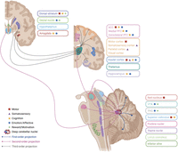
The cerebellum sends efferent projections to cortical and subcortical regions involved in a multitude of functions. Purkinje cell axons synapse in the deep cerebellar nuclei to form first-order projections. Second-order projections are formed by neurons in the deep cerebellar nuclei, which project to the thalamus and various subcortical regions. Third-order projections terminate in the cerebral cortex and basal ganglia. Based on Amino et al. (2001), Asanuma et al. (1983), Baek et al. (2022), Carta et al. (2019), Dietrichs et al. (1985), Faull and Carman (1978), Frontera et al. (2020), Haines and Dietrichs (1984), Ichinohe et al. (2000), Kelly et al. (2020), Kelly and Strick (2003), Pisano et al. (2021), Schmahmann (1996), Wagner et al. (2021), and Xiao et al. (2018). ACC, anterior cingulate cortex; PFC, prefrontal cortex; VTA, ventral tegmental area; PAG, periaqueductal gray. Created with BioRender.com.
Interestingly, stimulation of the motor cortex shows therapeutic efficacy in chronic pain syndromes, with evidence that its mechanism of analgesic action arises from antidromic recruitment of thalamocortical pathways to engage additional cortical and brainstem structures (Peyron et al., 2007; Antal et al., 2010; Nguyen et al., 2011; DosSantos et al., 2016). Motor cortex stimulation also modulates affective dimensions of pain, presumably via its interactions with regions such as the ACC and insular cortex (Picarelli et al., 2010; DosSantos et al., 2016). Considering these findings, a role for the cerebellum in guiding pain-related motor and/or affective responses is worth further exploration.
Parts of the medial PFC also receive efferent outputs from lobules IV–VI and Crus I of the cerebellum. Lobules IV and V project to the ACC, while vermal lobule VI and Crus I exhibit dense projections to the prelimbic and orbitofrontal cortices (Kelly et al., 2020; Pisano et al., 2021). The medial PFC is involved in numerous higher-order functions, such as integrating memories from prior experiences to guide goal-directed behavior, decision-making, and emotional responses (Etkin et al., 2011; Euston et al., 2012). Its role in many affective and cognitive components of pain such as those related to descending modulation, cognitive ruminations of pain, and psychiatric comorbidities of depression and anxiety have been well documented across human and animal studies (Metz et al., 2009; Bushnell et al., 2013; Kucyi et al., 2014; Kummer et al., 2020; Crawford et al., 2023a). Therefore, cerebellar activation in pain might also represent affective and cognitive dimensions of pain processing.
Structures involved in processing reward and motivation are also targets of cerebellar regions that are frequently associated with pain. Lobule VI sends output to the nucleus accumbens (NAc), while Purkinje cells in Crus I project to the VTA via the dentate nucleus (Pisano et al., 2021; Baek et al., 2022). The NAc and VTA are nodes of the pain matrix, so further interrogation into whether the cerebellum might exert a modulatory influence over motivational aspects of pain processing is warranted. Additionally, chemogenetic activation of Crus I Purkinje cells, that were previously identified to project to the VTA via the dentate nucleus, reduced depressive-like behaviors in mice that had been chronically stressed (Baek et al., 2022). This illustrates the potential for cerebellar activity to influence affective behavioral states, and in light of this, a cerebellar role in certain affective comorbidities of pain is also possible.
Further reason for contemplating the cerebellar involvement in affective and cognitive dimensions of pain is grounded in its communication with the hippocampus (Watson et al., 2019). Vermal lobule VI and Crus I project into the hippocampal dentate gyrus via the fastigial and dentate nuclei, respectively (Watson et al., 2019). Additionally, optogenetic stimulation of vermal lobules IV and V, as well as hemispheric lobule VI, increased hippocampal neuronal activation and interfered with performance on a hippocampal-dependent spatial memory task (Zeidler et al., 2020). The hippocampus has been proposed to process affective components of pain, with hippocampal abnormalities present in animal models of neuropathic pain and in patients with complex regional pain syndrome and chronic low-back pain (Mutso et al., 2012). In a spared nerve injury model of neuropathic pain, mice showed deficits in the memory-dependent contextual extinction of learned fear but not cued extinction (Mutso et al., 2012). The contextual extinction of conditioned fear is a hippocampus-dependent phenomenon, while cued extinction is hippocampus independent, so it was suggested that the experience of ongoing pain impaired hippocampal function, as corroborated by altered hippocampal neurogenesis and synaptic plasticity exhibited by these mice (Mutso et al., 2012). Given these hippocampal associations with pain, it would be worthwhile to explore whether cerebellar communications with the hippocampus are related to its involvement in pain.
Potential roles for cerebellar regions in acute pain processing
Motor control for defensive behaviors
As described earlier, the primary function of acute sensations of pain is to warn the individual of danger and to produce appropriate defensive responses. Given this, along with the historical view of the cerebellum as a comparator to refine ongoing movements, one might speculate that the cerebellum is critical for efficient expression of defensive behaviors in response to painful stimuli (Eccles et al., 1967; da Silva et al., 2023). Indeed, it has been hypothesized that the cerebellum regulates the timing of movements to enable coordinated behavior and motor learning, which would be critical for effective defensive behaviors (Spencer and Ivry, 2013; Chéron et al., 2016). Although changes in cerebellar activity during acute pain may be involved in modifying movements, participants in neuroimaging studies are generally instructed to inhibit movements to prevent movement-related artifacts. Given this, many have speculated that changes in cerebellar blood oxygen level dependent (BOLD) signals during acute pain paradigms represent inhibition of defensive behaviors. Consistent with this idea, experimental animal studies have shown that ventrolateral PAG stimulation, which regulates passive coping behaviors in response to pain such as quiescence, can directly inhibit nociceptive input to the cerebellum (Cerminara et al., 2009). Therefore, it is possible that this communication between the PAG and cerebellum is involved during the inhibition of behavioral responses in human imaging studies.
Several lines of evidence argue against the hypothesis that the cerebellum is exclusively involved in modulating motor output related to pain, however. First, sex differences showing that males were the main contributors to cerebellar activation during acute muscle and cutaneous pain makes this hypothesis unlikely (Henderson et al., 2008). Second, it has been shown that noxious heat can produce cerebellar activation even when participants are under general anesthesia, with activation magnitudes being similar to those when the participants were not sedated (Hofbauer et al., 2004). Third, cerebellar regions for motor control are thought to be distinct from those involved in affective and cognitive processes, as suggested by studies in patients with cerebellar lesions (Stoodley and Schmahmann, 2010; Stoodley et al., 2016). Generally, damage to anterior regions, typically lobules IV–V and extending into lobule VI, are associated with motor deficits without cognitive-affective impairments, while damage to posterior regions including lobules VII–VIII, Crus I, and Crus II result in cerebellar cognitive affective syndrome (Stoodley et al., 2016). Accordingly, lesions to both anterior and posterior regions led to motor and cognitive-affective deficits. Given these findings, it is possible that cerebellar activation during acute noxious stimuli is related not only to defensive behavioral expression, but also to another aspect of the pain experience.
Pain anticipation
Some studies report that cerebellar activation during experimental pain is related to the anticipation of pain, rather than the experience of pain per se (Ploghaus et al., 1999, 2000). Using fMRI in healthy participants exposed to noxious cutaneous heat, Ploghaus et al. (1999) demonstrated that cerebellar activation in regions responsive to the experience of pain were distinct from cerebellar regions activated in anticipation of pain. In contrast, some fMRI studies have reported that cerebellar activation in response to expected pain overlaps with cerebellar regions activated during the experience of pain (Koyama et al., 2005; Welman et al., 2018). These opposing accounts may be explained by discrepancies in the modalities of noxious stimulation and paradigms used for cuing noxious stimulation. Nevertheless, several studies have documented an association between cerebellar activity and the expectation of pain (Ploghaus et al., 1999; Koyama et al., 2005; Welman et al., 2018).
The cerebellum has been regarded as a site that encodes discrepancies between expected outcomes and established outcomes, otherwise known as prediction errors (Popa and Ebner, 2018). While the encoding of prediction signals has traditionally been applied to motor processing (Wolpert et al., 1998), it is also relevant to pain (Ploghaus et al., 2000), sensory processing (Schlerf et al., 2012), temporal processing (Reilly et al., 2008; Roth et al., 2013), language (Lesage et al., 2017), and conditioned fear (Ernst et al., 2019). In healthy participants, increased fMRI activation of the cerebellum was reported when noxious heat stimulation was unexpectedly omitted (Ploghaus et al., 2000) and when expectations of thermal pain intensity were incorrectly cued, thereby differing to what was expected (Zeidan et al., 2015b). Consistent with this, we recently reported that when the stimulus intensities did not match expectations, various brain areas including the ACC, dorsolateral PFC, PAG, and cerebellar lobules V and VI displayed significantly different patterns of activity compared to instances when stimulus intensity and pain expectations were matched (Henderson et al., 2020). These data implicate the cerebellum in detecting mismatches between actual and anticipated pain, although one should bear in mind that the expectation of pain and the experience of pain are not necessarily processed as mutually exclusive events, so further investigations are needed.
Attentional regulation of noxious stimuli
Our attention to noxious stimuli contributes extensively to our experience of pain (Villemure and Bushnell, 2002; Legrain et al., 2009). For example, distraction from noxious stimuli leads to reductions in the reported intensity and unpleasantness of pain (Bantick et al., 2002; Quiton and Greenspan, 2007; Boyle et al., 2008), whereas focusing attention on noxious stimuli can have different effects, either increasing the intensity and unpleasantness of pain or increasing one's tolerance to pain (Ahles et al., 1983; Miron et al., 1989). While studies have implicated regions such as the ACC, orbitofrontal cortex, and insular cortex in attentional states and pain (Bantick et al., 2002; Brooks et al., 2002), few studies have investigated contributions from the cerebellum. This is surprising given the emerging role of the cerebellum in attentional disorders such as attention-deficit hyperactivity disorder (Castellanos et al., 1996, 2002; Berquin et al., 1998; Bledsoe et al., 2011; Stoodley, 2014). Furthermore in humans, it has been shown that cerebellar lesions to the hemispheric lobules or nuclei impair performance on various attentional tasks (Courchesne et al., 1994; Gottwald et al., 2003) and inhibition or excitation of cerebellar sites can, respectively, interfere with or improve attention (Arasanz et al., 2012; Esterman et al., 2017; Mannarelli et al., 2019). Importantly, cerebellar activation measured via fMRI during a visual attention task without motor requirements was distinct from activation measured during the same attention task when a motor response was required. This suggests that the cerebellar sites involved in attention are unrelated to motor activity (Allen et al., 1997).
In recognition of the evidence for the modulation of attention by the cerebellum, it seems reasonable to consider how the cerebellum may be involved in directing attentional states in the context of noxious stimulation, for example, via its efferent projections to attention-regulating sites such as the ACC or the PFC. To our knowledge, no studies have directly examined this, although an fMRI study of healthy participants who received a mechanical pinprick to the hand found increased functional connectivity between a pain-activated cerebellar cluster and cortical regions associated with attentional function, such as the dorsolateral PFC (Diano et al., 2016). Indirectly, this implies the cerebellum modifies its connections with cortical regions known to be involved in attention. More direct insights into this role could be acquired by exploring how functional connectivity is altered under conditions in which attention is focused toward or away from noxious stimulation. In summary, future studies should reflect on whether cerebellar activation during noxious stimulation has a role in guiding attention related to the experience of pain.
Regulation of emotional states during noxious stimulation
Frequently discussed in parallel with the attentional regulation of pain are the emotional processes of pain modulation, although the neural processes underlying attentional and emotional modulation are distinct (Villemure et al., 2003; Villemure and Bushnell, 2009; Bushnell et al., 2013). The interaction between pain and emotional states and/or mood has been thoroughly documented (Villemure and Bushnell, 2002). Typically, the induction of negative moods increases the unpleasantness and intensity of pain (Rainville et al., 2005; Kenntner-Mabiala et al., 2008; Loggia et al., 2008), whereas the induction of positive mood is associated with increases in pain tolerance (Zelman et al., 1991; Weisenberg et al., 1998; de Wied and Verbaten, 2001). These effects are thought to arise from the capacity for emotional states to engage sites of the pain matrix, including the PFC, ACC, insular cortex, amygdala, and PAG. Activation or suppression of these regions can influence the recruitment of descending modulatory pathways (Willis and Westlund, 1997; Ossipov et al., 2014).
It is important to note that the vermal and hemispheric regions of the posterior cerebellum are consistently activated in human neuroimaging studies by experimental inductions of various emotions (Reiman et al., 1997; Paradiso et al., 1999; Liotti et al., 2000; Baumann and Mattingley, 2012). In addition, patients with cerebellar lesions often present with disturbances to affective processing (Schmahmann and Sherman, 1998; Turner et al., 2007). Moreover, repetitive transcranial magnetic stimulation (TMS) of the cerebellum can either elevate or depress mood (Schutter et al., 2003; Schutter and van Honk, 2009). Despite this evidence, studies to date have neglected to investigate cerebellar involvement in the emotional regulation of acute pain. Several studies have used fMRI to investigate neural correlates underlying experimentally induced mood on pain modulation (Wagner et al., 2009; Yoshino et al., 2010; Zunhammer et al., 2016; Jo et al., 2021) yet the function of the cerebellum is seldom explored. In a study that assessed cutaneous thermal pain thresholds and cold pain tolerance in pediatric patients with cerebellar lesions, patients with lesions encompassing Crus I and Crus II displayed reductions in their cold pain tolerance, while no differences in cold pain thresholds were detected (Silva et al., 2019). Therefore, it was proposed that the posterior cerebellum is involved in the affective processes of pain, rather than the sensory discrimination aspect. Moreover, patients also demonstrated weaker fMRI activation in the dorsolateral PFC and insular cortex during painful heat stimulation, implying that in the absence of an intact posterior cerebellum, there is atypical engagement of these cortical structures involved in affective pain processing (Silva et al., 2019). While this study lends relevant insights to how the cerebellum may contribute to the affective dimension of pain processing, comparable studies examining pain in patients with cerebellar lesions or ataxias are scarce. To our knowledge, only one analogous study exists (Ruscheweyh et al., 2014), as will be discussed in the later section, Regulation of descending modulatory pathways. Certainly, more detailed examination of whether cerebellar activation and lesions are associated with the regulation of emotional processes in acute pain would generate many stimulating insights.
Encoding reward processes associated with pain
The aversive nature of pain has been described as a punishment, which gives it motivational properties, because the relief of pain is a rewarding outcome (Navratilova et al., 2012; Nees and Becker, 2018). Cumulative evidence demonstrates an overlap between the neural processing of pain and that of reward. Indeed, many regions of pain are also implicated in reward processing, including the amygdala, insular cortex, ACC, PFC, striatum, and NAc (Baliki et al., 2010; Becerra et al., 2013; Nees and Becker, 2018). Moreover, it has been shown that dopaminergic and opioid signaling in the NAc is associated with placebo analgesia in healthy participants, and that pain relief operates as a reward, engaging dopaminergic signaling in mesolimbic reward circuits involving the VTA and NAc (Scott et al., 2008; Navratilova et al., 2012).
Recent investigations built upon neuroimaging studies (Holstege and Georgiadis, 2004; Tanaka et al., 2004) have demonstrated that the cerebellum is involved in reward processing. Various facets of reward signaling are processed by cerebellar granule cells (Wagner et al., 2017) and climbing fibers in animals (Heffley and Hull, 2019; Kostadinov et al., 2019; Larry et al., 2019). Moreover, a direct monosynaptic excitatory projection from the deep cerebellar nuclei into the VTA was identified in mice, and optogenetic stimulation of this projection was behaviorally rewarding, as shown by increased preference for the zone in the testing arena associated with stimulation (Carta et al., 2019). Lastly, a recent meta-analysis of fMRI investigations of reward-based tasks indicated that reward outcome was associated with activation in cerebellar lobule VI, while reward anticipation was associated with activation in bilateral lobules V–VI, posterior vermis, and left Crus I (Kruithof et al., 2023). Importantly, several of these regions overlap with cerebellar regions implicated in pain processing (Moulton et al., 2010; Alshelh et al., 2016).
In light of this, it seems natural to ask whether cerebellar activity during noxious stimulation is related to reward processing. Moulton et al. (2011) put forward a similar idea, in exploring whether fMRI activation in the cerebellum observed during noxious heat stimulation was associated with the encoding of aversive stimuli. Aversive stimuli presented as noxious cutaneous heat and the viewing of unpleasant images resulted in separate activation patterns in bilateral regions of the posterior cerebellum, as well as a group of overlapping activated regions encompassing lobule VI, Crus I, and lobule VIIb of the left hemisphere (Moulton et al., 2011). Regions of cerebellar activation for noxious heat were found to be positively correlated with activation in higher regions typically involved in the sensorimotor and affective domains of pain, such as the motor and somatosensory cortices, thalamus, putamen, dorsolateral PFC, insular cortex, and PAG. In contrast, the overlapping cerebellar regions activated by both noxious heat and unpleasant images were negatively correlated with limbic areas of the subgenual ACC, hypothalamus, and parahippocampal gyrus. These results suggest the presence of a distinct cerebrocerebellar pathway that encodes aversive stimuli, including pain, to ultimately guide motivational behavior (Moulton et al., 2011, 2014). This provides reasonable support for the hypothesis that the cerebellum processes reward and aversion during pain. Additional studies, including ones using animal models of pain, could provide insight into whether this role requires engagement with traditional mesolimbic and mesocortical reward pathways involving the VTA, NAc, and PFC.
Potential roles for cerebellar regions in chronic pain processing
Participation in somatosensory processing
In addition to the persistent perception of pain, chronic pain often presents with hyperalgesia and/or allodynia (Treede et al., 2019). Hyperalgesia results from a reduction in nociceptive threshold to produce an increased pain response, and allodynia arises when pain is perceived to a stimulus that was previously innocuous (Loeser and Treede, 2008). Given that the cerebellum encodes somatosensory inputs (Grodd et al., 2001), cerebellar changes in chronic pain may reflect the sensory discrimination of noxious inputs or adaptive changes to sensory inputs (Kim et al., 2015). Whereas cerebellar representations of innocuous somatosensory inputs to the foot and hand appear to be largely unilateral (Grodd et al., 2001; Buckner et al., 2011; Wiestler et al., 2011), thermal, mechanical, and electrical nociceptive stimulation of the arm, leg, face, fingers, or toes activates bilateral cerebellar regions (Coghill et al., 2001; Dimitrova et al., 2003; Borsook et al., 2008; Moulton et al., 2010; Welman et al., 2018). In agreement with this, the meta-analysis by Moulton et al. (2010) revealed that most neuroimaging investigations of experimental pain have found bilateral activation in the cerebellum. Additionally, numerous studies have reported cerebellar activation only ipsilateral to the site of noxious stimulation, few showed contralateral-only activation. Similarly, the majority of studies of pathological chronic pain reported ipsilateral-only or bilateral cerebellar activation (Moulton et al., 2010). It is unclear why these different activation patterns exist, but it is interesting to note a meta-analysis of neuroimaging studies that documented cerebellar activation during various tasks (Stoodley and Schmahmann, 2009). This analysis found that bilateral cerebellar activation was typically associated with executive and emotional tasks, whereas sensorimotor tasks predominantly engaged unilateral sites (Stoodley and Schmahmann, 2009). In view of this, it may be reasoned that bilateral cerebellar engagement in chronic pain results from global cortical interactions, reflecting cognitive and affective elements of the nociceptive response, such as pain unpleasantness. On the other hand, the somatosensory discrimination of pain, such as pain intensity, may be concerned to a larger extent with unilateral cerebellar activation. The following sections will examine additional facets of chronic pain, supplementary to somatosensory discrimination, that may contribute to the cerebellar activation observed across neuroimaging studies.
Regulation of descending modulatory pathways
Symptoms of hyperalgesia and allodynia are thought to develop from pathophysiological processes such as central sensitization, where nociception becomes hyperexcitable in the central nervous system. A common explanation for how central sensitization arises points to the suppression of descending modulatory pathways (Kwon et al., 2014). Given the anatomical connections between the cerebellum and regions of nociceptive modulation, it is worth considering whether cerebellar efferent projections might exacerbate central sensitization processes by influencing pathways that modulate nociception. This idea was previously suggested after it was shown that electrical stimulation of the left anterior cerebellar cortex in rats significantly dampened responses in dorsal horn neurons to noxious pressure and pinch (Hagains et al., 2011). Specifically, it was proposed that stimulation of the cerebellar cortex diminished inhibitory outputs from the emboliform nucleus, leading to disinhibition of brainstem regions involved in descending modulation and consequently, increased descending inhibitory modulation. This notion is supported by the finding that patients with cerebellar infarcts display weaker placebo analgesia and offset analgesia, which describes a disproportionately large pain reduction in response to a small decrease in noxious intensity, therefore suggesting impaired recruitment of endogenous inhibitory nociception (Ruscheweyh et al., 2014). Similarly, inhibitory transcranial direct current stimulation (tDCS) or excitatory tDCS over the cerebellum respectively reduced or enhanced endogenous inhibition in healthy participants, although the effects were not large enough to be deemed clinically relevant (Stacheneder et al., 2022). While in recent fMRI studies we found that placebo analgesia was not associated with cerebellar activation (Crawford et al., 2023b), we have shown that conditioned pain modulation is associated with lobule V/VI activation (Youssef et al., 2016). Despite the available evidence suggesting that cerebellar activation in chronic pain may relate to inhibitory pain modulation, much uncertainty remains as to which regions and pathways of the cerebellum and brainstem are precisely involved, so further work is necessary to confirm this relationship.
Engagement in atypical salience processing
A relatively modern explanation for chronic pain invokes the concept of aberrant salience processing (Borsook et al., 2013). Salience processing involves the integration of internal and external information from various sensory, visceral, and autonomic inputs, such that a rank of homostatic importance can be assigned to this information to subsequently guide behavior (Seeley et al., 2007). When exposed to noxious stimulation, the salience that we attribute to the resulting aversive state drives our inclination to seek out a balanced homostatic state. Seeley et al. (2007) described a system of interacting neuroanatomical regions involved in salience processing, which is typically referred to as the salience network. The network is largely localized to the dorsal ACC and anterior insular cortex, but also encompasses sites in the PFC, amygdala, hypothalamus, VTA, and thalamus (Seeley et al., 2007). Importantly, cerebellar nodes of the salience network have been identified, specifically lobules VI, VIIB, Crus I, Crus II, and the dentate nucleus (Habas et al., 2009), which coincidentally overlap with cerebellar sites corresponding to pain. Owing to its considerable connectivity with previously-identified nodes of the salience network, the cerebellum may be a significant contributor to salience processing (Sang et al., 2012).
Notably, it has been suggested that in chronic pain patients, baseline salience function becomes sensitized to pain-relevant stimuli, and moreover, that patients present with abnormalities in assigning salience to external and internal stimuli (Borsook et al., 2013). Corroborating this, abnormalities in salience network connectivity have been reported in patients with chronic widespread pain (van Ettinger-Veenstra et al., 2019), chronic low back pain (Kim et al., 2019), fibromyalgia (Pinto et al., 2022), trigeminal neuralgia (Xu et al., 2022), irritable bowel syndrome (Icenhour et al., 2017), and migraine (Veréb et al., 2020).
With this in mind, we asked whether cerebellar activation within the context of chronic pain may reflect disruptions to salience processing. In an fMRI study comparing 135 patients with chronic low-back pain to healthy controls, patients demonstrated increased functional connectivity between the salience network and the cerebellum, specifically with lobules VI and Crus I, at rest. This suggests that baseline salience network function is altered in chronic pain patients (Kim et al., 2019). However, the bulk of studies that explore salience network activation in chronic pain patients lack any detailed analysis of cerebellar activation patterns, so our understanding into this relationship is limited. As such, future studies addressing this would prove highly insightful.
Pain-related fear processing
The fear avoidance model of chronic pain postulates that avoidance of movements or daily activities are driven by one's fear of pain and anticipated pain-related outcomes (Vlaeyen and Linton, 2000). Relief from this fear and anticipated pain is achieved by engagement of these avoidant behaviors. This results in a maladaptive learning response in which avoidant behaviors are repeated, thereby exacerbating chronic pain (Vlaeyen and Linton, 2000; Vlaeyen, 2015). Improved insights into the underlying neural processes of pain-related fear may assist with the correction of these maladaptive avoidant behaviors in chronic pain.
Considering a role for the cerebellum in processing pain-related fear is worthwhile, given the importance of the cerebellum in conditioned fear. Animal studies show that lesions to the cerebellar vermis, specifically to vermal lobules IV–VI, interfere with the acquisition and retention of a conditioned fear response (Supple and Leaton, 1990; Sebastiani et al., 1992; Supple and Kapp, 1993). Moreover, the vermis projects to the cerebellar fastigial nucleus, which consists of efferents to the ventrolateral PAG that regulate the strength of conditioned fear responses (Frontera et al., 2020). Furthermore, patients with vermal lesions lacked the bradycardia response associated with conditioned fear that was displayed in healthy controls (Maschke et al., 2002). Despite the cerebellar contributions to conditioned fear, few have directly considered whether its activation in pain studies is relevant to pain-related fear.
To assess neural activation associated with pain-related fear conditioning, Benson et al. (2014) examined neural fMRI responses during classical conditioning phases when neutral visual stimuli were paired with rectal distensions. They identified a cerebellar correlate, such that when the visceral-pain-related fear memory was reactivated after extinction, females showed greater cerebellar, hippocampal, and thalamic activation than males, despite there being no sex differences in pain threshold or intensity ratings. In contrast, Biggs et al. (2020) conducted a meta-analysis of MRI studies investigating pain-related conditioned fear and non-pain related fear and failed to note any cerebellar activation associated with pain-related fear. This may have been due to a paucity of detailed studies on the cerebellum. Thus, there is insufficient evidence to determine whether cerebellar activity in chronic pain populations is associated with pain-related fear processes.
Internal models within the cerebellum during pain processing
We have thus far explored various ways in which the cerebellum may be involved in pain processing via its connections to higher cortical areas, without discussing the recognized role of the cerebellum in generating internal models (Wolpert et al., 1998; Ito, 2005, 2008). Therefore, we will now briefly consider how cerebellar activation in pain may relate to internal models generated within the cerebellum.
Internal models are created by localized neural networks to represent systems or processes in the external world (Wolpert et al., 1995). For example, the cerebellar circuitry is thought to create internal models to predict how motor commands will affect movements of body parts. These models are thought to be refined through a continual learning process as movements are reiterated (Wolpert et al., 1998; Ito, 2005). In accordance with the recent literature supporting a role for the cerebellum in non-motor domains (Gatti et al., 2021; Wu et al., 2022; Frontera et al., 2023; Kruithof et al., 2023), the cerebellum has also been proposed to generate internal models for mental processes, such as memory (Ito, 2008). While it is beyond the scope of this review to describe the computational systems of these internal models at length, these internal models are believed to facilitate the production of accurate movements without the need for immediate sensory feedback from the external world, by directly communicating with the sensorimotor effector regions. Moreover, cerebellar internal models may explain how implicit mental processes arise without the engagement of higher cortical regions, thus bypassing conscious awareness (Ito, 2008).
Does cerebellar activation documented in pain studies represent feedback integration and learning within internal model systems, rather than the experience of pain per se? For any given body part that receives noxious stimulation, activation in the cerebellum may symbolize the integration of sensorimotor signals in the internal model of said body part, which facilitates the adaptation of an existing internal model to a setting of noxious stimulation. This theory may explain activation of vermal lobules IV–V and hemispheric lobules IV–VI during noxious mechanical stimulation to the hand, given that these regions also exhibit functional connectivity with sensorimotor cortical regions (Diano et al., 2016). This coordinated activity may result from interactions between the internal model and the sensorimotor effector sites. If this is the case, cerebellar activation may arise in any context of sensorimotor stimulation that leads to adaptation in internal models. In other words, cerebellar activation may not relate specifically to pain processing, but rather to a unique consequence of pain processing, such as sensorimotor engagement. However, this theory does not account for the considerable sex differences in cerebellar activation documented during experimental cutaneous and muscle pain, as alluded to earlier (Henderson et al., 2008).
Alternatively, cerebellar activation in pain may represent engagement of internal models within the cerebellum pertinent to cognitive processes recruited for the experience of pain. Cerebellar internal models for mental processes are believed to engage with higher order regions including the PFC, ACC, and hippocampus to perform and refine mental tasks (Ito, 2008). For example, given that the experience of pain demands the recruitment of attentional processes, cerebellar activation could simply represent the convergence of feedback on an internal model for attention, which is not necessarily exclusive to pain. Consistent with this, noxious mechanical stimulation to the hand activated a cluster comprising hemispheric lobule VI, Crus I, and Crus II, which was functionally connected with sites involved in attention, such as the dorsolateral PFC, and cingulate cortex (Diano et al., 2016).
Internal models of mental processes in the cerebellum may also be evaluated in the context of placebo analgesia. Placebo analgesia is a phenomenon which incorporates several intricate cognitive processes such as memory and expectation (Colloca et al., 2013). When participants are experimentally conditioned to expect pain relief from a placebo treatment, adaptation would not be required for the existing internal model during a placebo analgesia response, as such adaptation would have previously been incorporated in the model throughout conditioning. Although this explains why Crawford et al. (2023b) did not identify cerebellar activation in placebo analgesia responders, a recent meta-analysis discordantly reported that placebo analgesia responses were negatively correlated with activation in lobule VI (Zunhammer et al., 2021).
Collectively, a vast array of sensorimotor, cognitive, and emotional processes exhibit cerebellar engagement independently of cerebellar engagement during noxious stimulation. Therefore, it is reasonable to reflect on whether cerebellar activation observed in pain may represent engagement of internal models of sensorimotor and mental processes emanating from the experience of pain. However, the conflicting evidence indicates the need for further investigation to specifically test this proposition. In addition, although internal models of emotion have not been extensively documented (Adamaszek et al., 2017; Petrosini et al., 2022), a further question is whether the cerebellum formulates internal models for emotional processes derived from the experience of pain, alongside cognitive and sensorimotor processes.
Further considerations regarding neuroimaging during pain
Thus far, we have explored what the reported changes to cerebellar activation detected by fMRI in pain studies could conceptually or functionally represent in pain processing, but it is also critical that we ask what cerebellar activation represents at the cellular level. Over 20 years ago, Logothetis (2002) compared local field potentials and single- and multi-unit activity with BOLD fMRI responses recorded simultaneously in the monkey visual cortex. BOLD fMRI responses were significantly correlated with local field potentials, but not single- and multi-unit activity, suggesting that BOLD fMRI signals primarily represent inputs and local processing, rather than spiking output. Moreover, it has been recently reported that local field potentials, which represent summed local synaptic activity, largely reflect inhibitory neuron activity (Teleńczuk et al., 2017), and that activity of inhibitory neurons, rather than excitatory neurons, is the primary driver of BOLD responses (Vazquez et al., 2018). Considering all this, cerebellar fMRI signals evoked in pain studies might represent local synaptic activity at Purkinje cells. Such activity likely encompasses the summation of excitatory inputs from ascending climbing and parallel fibers, as well as local inhibitory interneuron activity.
Since Purkinje cells project to the deep cerebellar nuclei, one would expect there to be concurrent changes in fMRI signals within the cerebellar cortex and deep cerebellar nuclei. However, we know of no study that has reported signal changes in deep cerebellar nuclei during noxious stimulation in humans. This is likely a consequence of the limited spatial acuity afforded by standard high-field fMRI, and its restricted capacity to detect the small cerebellar nuclei. However, the recent advent of ultra-high-field fMRI will allow investigation into smaller brain regions, including the deep cerebellar nuclei, in response to noxious stimulation. Such investigations will be pivotal in our understanding of the basis of cerebellar activation in pain.
Therapeutic potential of the cerebellum
The evidence supporting the hypotheses presented in this review is modest or largely indirect. Without question, however, thorough investigation into the relationships between the cerebellum and pain, particularly chronic pain, will provide beneficial insights into the pathophysiological mechanisms driving chronification of pain. Notably, many of the current pharmacological therapeutics for chronic pain have limited efficacy and are ill-suited for long-term use. Therefore, alternative therapies are needed. Toward this goal, we end this review by considering how the cerebellum may be targeted with non-pharmacological therapies to reduce the burden of chronic pain.
Transcranial direct current stimulation
tDCS has emerged as a promising therapeutic tool for various medical conditions including movement disorders, Parkinson's disease, Alzheimer's disease, psychiatric disorders, and chronic pain (Fregni et al., 2015; Lefaucheur et al., 2017; Pinto et al., 2018). With respect to chronic pain, most studies use anodal tDCS over the primary motor cortex, although stimulation of the dorsolateral PFC and primary visual cortex is sometimes adopted [as reviewed in Lefaucheur et al. (2017); Pacheco-Barrios et al. (2020)]. Despite reports of significant improvements to pain following tDCS in various chronic pain conditions, its clinical recommendation for therapeutic use remains restricted, due to study limitations relating to methodology and sample size (Lefaucheur et al., 2017).
Interestingly, Bocci et al. (2015) showed that cerebellar tDCS in healthy controls reduced the perception of cutaneous thermal pain evoked with infrared laser potentials, possibly by altering cerebellar engagement with somatosensory and cingulate cortices. Furthermore, in patients with phantom limb pain, anodal tDCS over the cerebellum significantly alleviated paroxysmal pain and nonpainful sensations in the phantom limb, although it had no effect on phantom limb pain and stump pain (Bocci et al., 2019). Although some evidence exists for a beneficial effect of cerebellar tDCS, more comprehensive studies are required to confirm this, and to investigate the specific therapeutic mechanisms involved.
Given that the cerebellum projects globally, including to numerous nociceptive cortical and subcortical sites, cerebellar tDCS may substantially affect interactions between regions. Such effects could be particularly relevant for targeting the regions implicated in elements of the pain experience discussed in this review, including the PFC, ACC, and nodes of the salience network. Indeed, it would be valuable for future cerebellar tDCS studies to additionally examine associated changes in nociceptive regions.
Studies have also suggested tDCS as a strategy to prevent the development of chronic pain (Spezia Adachi et al., 2012; Fregni et al., 2018; Pacheco-Barrios et al., 2020). In rats exposed to chronic restraint stress, which subsequently led to thermal hyperalgesia and mechanical allodynia, repetitive anodal tDCS delivered at the conclusion of the stress exposure reversed stress-induced pain behaviors (Spezia Adachi et al., 2012). Additionally, tDCS administered prior to chronic stress exposure in rats prevented stress-induced hyperalgesia (Fregni et al., 2018). Although it appears that tDCS could be useful as an early therapeutic intervention to mitigate chronic pain development, further research extending into clinical samples is required, both to corroborate this hypothesis and to elucidate the specific mechanisms and outcomes of cerebellar tDCS.
Mindfulness meditation
An alternative to directly targeting the therapeutic potential of the cerebellum via tDCS is to facilitate its recruitment indirectly using cognitive or behavioral approaches such as those involving mindfulness. Although recent investigations have detailed beneficial effects of mindfulness meditation in reducing experimental pain and various chronic pain pathologies (Grossman et al., 2007; Fox et al., 2011; Gaylord et al., 2011; Zeidan et al., 2011, 2015a; Izgu et al., 2020), meta-analyses have reported small effect sizes, and its mechanisms of efficacy in pain require clarification (Chiesa and Serretti, 2011; Hilton et al., 2017).
Studies of the neuroanatomical substrates of mindfulness-induced analgesia suggest contributions from numerous cognitive and emotion-processing regions including the cerebellum, as well as the dorsolateral PFC, orbitofrontal cortex, insular cortex, ACC, and thalamus (Zeidan et al., 2011; Riegner et al., 2022). While many of these structures have been discussed in the context of mindfulness, details regarding the cerebellum remain sparse (Zeidan et al., 2011; Nascimento et al., 2018). Notably, mindfulness-based interventions induced changes to gray matter volume in the cerebellar vermis and posterior hemispheric lobules, and altered cerebellar functional connectivity with higher cortical structures, such as the orbitofrontal cortex and putamen (Hölzel et al., 2011; Santarnecchi et al., 2021), but a large randomized control trial recently found no gray matter volume changes in the cerebellum following a similar intervention (Kral et al., 2022). Mindfulness-based interventions may instead engage the cerebellum via its influence on the salience network (Kilpatrick et al., 2011; Hasenkamp and Barsalou, 2012; Bremer et al., 2022). With this said, it is uncertain as to what extent these cerebellar changes relate to the analgesic effects of mindfulness-based interventions, and substantial insights are needed to advocate for its applicability in the clinic.
Conclusions
Accumulated evidence from human and animal studies of pain has reliably suggested a function for the cerebellum in pain, yet investigations into what exactly these functions may be are still lacking. Given the diverse array of motor and non-motor functions of the cerebellum, we speculate that the cerebellum exerts a modulatory influence on multidimensional facets of pain processing by communicating with cortical and subcortical structures. We draw particular attention to how cerebellar lobules IV–VI and Crus I interact with higher-order structures, as these regions frequently exhibit associations with pain. Upon review of the anatomical literature, we note projections into sensorimotor regions, and additionally highlight connections with regions implicated in cognitive and emotional processing – notably the dorsolateral PFC, medial PFC, VTA, NAc, and hippocampus – all of which have been implicated in pain processing independently of cerebellar function. Based on these connections, we have proposed various mechanisms through which the cerebellum influences acute and chronic pain, with particular focus on emotional and cognitive processes. While the speculations put forward here have been addressed independently, we acknowledge that they need not act exclusively. Considering the domain-general function of the cerebellum, it holds vast therapeutic potential for modulating functions that may be significant to pain. For this reason, comprehensive examinations into the documented cerebellar associations with pain are much needed.
Footnotes
- Received September 3, 2023.
- Revision received March 6, 2024.
- Accepted March 6, 2024.
-
We thank the National Health and Medical Research Council (NHMRC).
-
The authors declare no competing financial interests.
- Correspondence should be addressed to Richelle Mychasiuk at Richelle.mychasiuk{at}monash.edu.
- Copyright © 2024 the authors
References
- ↵
- ↵
- ↵
- ↵
- ↵
- ↵
- ↵
- ↵
- ↵
- ↵
- ↵
- ↵
- ↵
- ↵
- ↵
- ↵
- ↵
- ↵
- ↵
- ↵
- ↵
- ↵
- ↵
- ↵
- ↵
- ↵
- ↵
- ↵
- ↵
- ↵
- ↵
- ↵
- ↵
- ↵
- ↵
- ↵
- ↵
- ↵
- ↵
- ↵
- ↵
- ↵
- ↵
- ↵
- ↵
- ↵
- ↵
- ↵
- ↵
- ↵
- ↵
- ↵
- ↵
- ↵
- ↵
- ↵
- ↵
- ↵
- ↵
- ↵
- ↵
- ↵
- ↵
- ↵
- ↵
- ↵
- ↵
- ↵
- ↵
- ↵
- ↵
- ↵
- ↵
- ↵
- ↵
- ↵
- ↵
- ↵
- ↵
- ↵
- ↵
- ↵
- ↵
- ↵
- ↵
- ↵
- ↵
- ↵
- ↵
- ↵
- ↵
- ↵
- ↵
- ↵
- ↵
- ↵
- ↵
- ↵
- ↵
- ↵
- ↵
- ↵
- ↵
- ↵
- ↵
- ↵
- ↵
- ↵
- ↵
- ↵
- ↵
- ↵
- ↵
- ↵
- ↵
- ↵
- ↵
- ↵
- ↵
- ↵
- ↵
- ↵
- ↵
- ↵
- ↵
- ↵
- ↵
- ↵
- ↵
- ↵
- ↵
- ↵
- ↵
- ↵
- ↵
- ↵
- ↵
- ↵
- ↵
- ↵
- ↵
- ↵
- ↵
- ↵
- ↵
- ↵
- ↵
- ↵
- ↵
- ↵
- ↵
- ↵
- ↵
- ↵
- ↵
- ↵
- ↵
- ↵
- ↵
- ↵
- ↵
- ↵
- ↵
- ↵
- ↵
- ↵
- ↵
- ↵
- ↵
- ↵
- ↵
- ↵
- ↵
- ↵
- ↵
- ↵
- ↵
- ↵
- ↵
- ↵
- ↵
- ↵
- ↵
- ↵
- ↵
- ↵
- ↵
- ↵
- ↵
- ↵
- ↵
- ↵
- ↵
- ↵
- ↵
- ↵
- ↵
- ↵
- ↵
- ↵
- ↵
- ↵
- ↵
- ↵
- ↵
- ↵
- ↵
- ↵
- ↵
- ↵
- ↵
- ↵
- ↵
- ↵
- ↵
- ↵
- ↵
- ↵
- ↵
- ↵
- ↵
- ↵
- ↵
- ↵
- ↵
- ↵
- ↵
- ↵
- ↵
- ↵
- ↵
- ↵
- ↵
- ↵
- ↵
- ↵
- ↵
- ↵
- ↵
- ↵
- ↵
- ↵
- ↵
- ↵
- ↵
- ↵
- ↵
- ↵
- ↵
- ↵
- ↵




