Cortical Tagged Synaptic Long-Term Depression in the Anterior Cingulate Cortex of Adult Mice
- 1Center for Neuron and Disease, Frontier Institute of Science and Technology, Xi’an Jiaotong University, Xi’an 710049, China
- 2Zhuomin Institute of Brain Research, Qingdao International Academician Park, Qingdao 266000, China
- 3CAS Key Laboratory of Brain Connectome and Manipulation, Interdisciplinary Center for Brain Information, The Brain Cognition and Brain Disease Institute, Shenzhen Institute of Advanced Technology, Chinese Academy of Sciences, Shenzhen 518055, China
- 4Department of Exercise & Health Science, Xi'an Physical Education University, Xi'an 710068, China
- 5Department of Physiology, Faculty of Medicine, University of Toronto, Toronto, Ontario M5S 1A8, Canada
-
Author contributions: X.-H.L. and M.Z. designed research; W.L. performed research; W.L. contributed unpublished reagents/analytic tools; W.L. analyzed data; W.L., Q.-Y.C., and Z.Z. wrote the paper.
Abstract
The anterior cingulate cortex (ACC) is a key cortical region for pain perception and emotion. Different forms of synaptic plasticity, including long-term potentiation (LTP) and long-term depression (LTD), have been reported in the ACC. Synaptic tagging of LTP plays an important role in hippocampus-related associative memory. In this study, we demonstrate that synaptic tagging of LTD is detected in the ACC of adult male and female mice. This form of tagged LTD requires the activation of metabotropic glutamate receptor subtype 1 (mGluR1). The induction of tagged LTD is time-related with the strongest tagged LTD appearing when the interval between two independent stimuli is 30 min. Inhibitors of mGluR1 blocked the induction of tagged LTD; however, blocking N-methyl-d-aspartate receptors did not affect the induction of tagged LTD. Nimodipine, an inhibitor of L-type voltage–gated calcium channels, also blocked tagged LTD. In an animal model of amputation, we found that tagged LTD was either reduced or completely blocked. Together with our previous report of tagged LTP in the ACC, this study strongly suggests that excitatory synapses in the adult ACC are highly plastic. The biphasic tagging of synaptic transmission provides a new form of heterosynaptic plasticity in the ACC which has functional and pathophysiological significance in phantom pain.
- anterior cingulate cortex
- long-term depression
- metabotropic glutamate receptor subtype 1
- multielectrode array
- synaptic tagging
Significance Statement
The anterior cingulate cortex (ACC) is a key cortical region for pain perception and chronic pain. Previous studies have reported a novel form of long-term heterosynaptic potentiation in the ACC. In this study, we discovered a long-term depression (LTD) form of synaptic tagging in the ACC of adult male and female mice. This form of tagged LTD requires the activation of metabotropic glutamate receptors. In an animal model of amputation, tagged LTD is reduced or completely blocked. Our results strongly suggest that brain cortices of adult mice are highly plastic and show biphasic tagging of plasticity. These findings of tagged LTD may provide a new direction for future treatment of phantom pain and amputation-related emotional disorders.
Introduction
Synaptic tagging and capture is a widely studied heterosynaptic form of plasticity associated with learning and memory in the hippocampus (Frey and Morris, 1997; P. Park et al., 2019; Bin Ibrahim et al., 2022). It involves two primary forms of long-term potentiation (LTP): early-phase LTP, which is induced by a weak tetanus stimulus and does not depend on protein synthesis, and late-phase LTP, which is induced by a strong tetanus stimulus and requires protein synthesis (Frey and Morris, 1997; Reymann and Frey, 2007; Bin Ibrahim et al., 2022). These two forms of LTP can interact heterosynaptically through synaptic tagging. This process begins with a strong stimulus that likely triggers the synthesis of plasticity-related proteins (PRPs), enhancing synaptic plasticity. The subsequent, or preceding weak stimulus, occurring within the decay time of the synaptic tag, establishes a synaptic “tag”. This tag captures a portion of these PRPs, leading to the establishment of a new LTP at the site of the weaker stimulus (Frey and Morris, 1997; Reymann and Frey, 2007; Nomoto and Inokuchi, 2018; P. Park et al., 2019). Synaptic tagging is proposed to play a key role in both learning and memory (Frey and Morris, 1997; Redondo et al., 2010; Rogerson et al., 2014).
The anterior cingulate cortex (ACC) is the frontal part of the cingulate cortex in mammalian brains and a key cortical region for chronic pain and emotion (Vogt, 2005; Zhuo, 2008, 2016b; Bliss et al., 2016). Hyperactivation of ACC is closely associated with many chronic pain conditions, which are always associated with fear, anxiety, and cognitive dysfunction (Frankland et al., 2004; Zhuo, 2008, 2016b; Koga et al., 2015b; Bliss et al., 2016). Different forms of LTP have been reported in the ACC (Bliss et al., 2016), and synaptic tagging of LTP has also been reported (M. G. Liu et al., 2018). In addition to LTPs, long-term depression (LTD) has been investigated in the ACC (Wei et al., 1999; Toyoda et al., 2005, 2007; S. J. Kang et al., 2012; Bliss et al., 2016). Low-frequency stimulation (LFS, 1 Hz for 15 min), which is reported to induce LTD in the hippocampus (Mulkey and Malenka, 1992; Dudek and Bear, 1993; Bear and Malenka, 1994; Bear and Abraham, 1996), has been found to induce reliable LTD in the ACC of adult mice (Wei et al., 1999; S. J. Kang et al., 2012; Bliss et al., 2016). Both N-methyl-d-aspartate (NMDA) receptor-dependent LTD and metabotropic glutamate receptor (mGluR)-dependent LTD have been reported in the ACC (Bliss et al., 2016; Koga et al., 2016; Xue et al., 2021).
Amputation causes long-lasting changes in the central nervous system and can cause phantom pain and limb sensation, as well as anxiety and depression (Katz and Melzack, 1990; Buonomano and Merzenich, 1998; Zhuo, 1998, 2012; Dostrovsky, 1999; Kaas, 2000; Flor, 2002; Bhutani et al., 2016; Fuchs et al., 2018; Aternali and Katz, 2019; Tsoulou et al., 2019). Previous studies found that digit amputation caused LTP-like responses in the ACC in vivo, prolonged synaptic activation, and increased the spontaneous neuronal activity of cortical pyramidal cells (Wei and Zhuo, 2001; Li et al., 2013). Activity-dependent immediate early genes such as c-Fos and the cyclic AMP-responsive element binding protein (CREB) also increased in the ACC and hippocampus after digit amputation (Wei et al., 1999, 2000). More interestingly, LFS failed to induce LTD after digit amputation in brain slices of adult mice (Wei et al., 1999; S. J. Kang et al., 2012). Recently, M. G. Liu et al. (2018) reported that synaptic tagging of LTP is blocked after tail amputation. Less is known about synaptic tagging of LTD and changes after digit/tail amputation. In the present study, by applying the multichannel electrode field recording, we successfully recorded tagged LTD in the ACC of adult mice, providing novel evidence that synaptic tagging can be biphasic in the cortex. We also found that the tagged LTD was blocked after amputation. Furthermore, inhibitors of mGluR1 blocked the induction of synaptic tagging of LTD. Together with our previous studies (M. G. Liu et al., 2018), our findings suggest that both tagged LTP and tagged LTD exist in synapses in the ACC of adult mice, and this frequency-dependent relation of LTP/LTD is altered after peripheral amputation.
Materials and Methods
Animals
C57BL/6 adult male/female mice were purchased from the Experimental Animal Center of Xi’an Jiaotong University, Charles River Laboratories, and Qingdao Qinda Animal Biotechnology (6–8 weeks old). All mice were kept in groups and maintained a 12 h light/dark cycle (lights turned on at 7 A.M.) in a storage room with a temperature of 23 ± 1°C and air humidity of 55–60%. Food and water were provided ad libitum. All experiments were carried out at Xi’an Jiaotong University, and the experimental protocol was approved by the Xi’an Jiaotong University Animal Protection and Use Committee, in accordance with the ethical guidelines of the Ethics Committee.
Preparation for brain slices of mice
The method of preparing brain slices of ACC was consistent with our previous methods (M. G. Liu et al., 2018; Zhou et al., 2023). Mice were anesthetized with isoflurane and killed by decapitation. The brain was quickly removed and placed into cold oxygenated (95% O2 and 5% CO2) artificial cerebrospinal fluid (ACSF) containing the following (in mM): 124 NaCl, 2.5 KCl, 1.0 NaH2PO4, 1 MgSO4, 2 CaCl2, 25 NaHCO3, 10 glucose, pH 7.35–7.45. The trimmed brain was glued to a microtome (Leica VT1200S) for coronal ACC slices (300 μm thickness per slice). The slices were then transferred to an oxygenated submerged recovery chamber containing ACSF solution (95% O2 and 5% CO2) at room temperature for at least 1 h.
Multichannel field potential recording
ACC extracellular multichannel field potential recording was performed using the MED64 channel recording system (Panasonic Alpha-Med Sciences). MED64 probe (P515A; 64 square planar microelectrode arrays arranged in 8 × 8 mode; interpolar distance, 150 μm; Panasonic Alpha-Med Sciences) requires a hydrophilic treatment before the experiment, with a method similar to that in our previous studies (see above, Preparation for brain slices of mice). Use 0.1% polyethyleneimine (Sigma-Aldrich) soaked in 25 mmol/L borate buffer, pH 8.4, and left at room temperature overnight. The probe surface is then carefully rinsed with sterile distilled water three times to remove possible harmful substances and avoid affecting the brain slices. Brain slices can be better attached to MED64 probes after hydrophilic treatment. The incubated brain slices were placed on the MED64 probe so that the different layers of ACC were covered by 64 electrodes as much as possible (Fig. 1A). After a slice position was fixed, the slice was carefully further and fully fixed with a mesh and an anchor (Warner Instruments, Harvard Apparatus) to ensure the stability of the slice during the experimental recording process. Throughout the experimental period, slices were continuously infused with fresh ACSF oxygenated (95% O2 and 5% CO2) at a rate of 2–3 ml/min with the help of a peristaltic pump (MINIPULS 3, Gilson).

A sample of synaptic tagging of LTD in the ACC of adult mice. A, Light microscopy photograph showing the location of an ACC slice on a MED64 probe (microelectrode size 50 × 50 μm, the interpolar distance of electrodes 150 μm). Electrical stimulation is separated into two distinct inputs. The S1 site is shown as a red circle for the strong LFS in the superficial layer; the S2 site is shown as a blue circle for the weak LFS in the deep layer. B, E–H, Individual data presentation of synaptic tagging recording in the ACC. Large arrows indicate the starting point of strong LFS application in the S1 site; small arrows mark the time point of tagged LFS delivery in the S2 site. B, The fEPSP slope demonstrates the induction of LTD by weak LFS if preceded, 30 min earlier, by a strong LFS (i.e., tagged LFS). Strong LFS-evoked LTD and tagged LFS-evoked tagged LTD last for at least 1 h in one channel of a slice. C, S1 and S2 are two relatively independent sites. Top, Representative traces indicated by numbers in the graph with calibration of channels that were activated by a paired pulse. Down, Bar histogram showing quantified data within the S1 and S2 sites. Left, Once at the S1 site and once at the S1 site (black)/once at the S2 site and once at the S1 site (red), in one slice. First pulse (blue) and second pulse (red). Right, Once at the S2 site and once at the S2 site (black)/once at the S1 site and once at the S2 site (red), in one slice. First pulse (green) and second pulse (purple); NS, no significance. D, Two mapped figures show the evoked field potentials in the ACC in the S1 site [left, field potentials were recorded from the other 63 channels 30 min before (black) and 30 min after (red) strong LFS being delivered to one channel marked as “s,” 45 channel in Layer Ⅲ] and S2 site [right, field potentials were recorded from the other 63 channels 30 min before (black) and 60 min after (red) weak LFS being delivered to one channel marked as “s,” 52 channel in Layer Ⅴ]. Superimposed sampled traces at different time points display two types of plasticity: channels exhibiting typical LTD are marked as (1), while channels without changes in the slope of the fEPSP are marked as (2). Star symbols indicate silencing responses. E, Individual data for channels showing LTD by the S1 input after receiving strong LFS 30 min postbaseline recording. In one slice analyzed, 13 channels consistently showed LTD. F, Individual data for tagged LTD; channels for the S2 input received tagged LFS 30 min after strong LFS. In one slice analyzed, 13 channels consistently showed tagged LTD. G, A summarized plot of the fEPSP slope demonstrates the induction of persistent LTD by strong LFS alone at the S1 site, lasting for 1.5 h across all channels in a slice. H, A summarized plot of the fEPSP slope shows the induction of LTD by weak LFS at the S2 site, and tagging of LTD persists for at least 1 h across all channels in a slice.
The slices are stored on the probe for at least 1 h before recording. A site was selected from 64 planar microelectrodes to test the stimulus, and a constant current pulse lasting 0.2 ms was given to the superficial layer (Layers Ⅱ–Ⅲ, for S1-strong LFS input) and deep layer (Layers Ⅴ–Ⅵ, for S2-tagged LFS input) of the ACC slices. The response was generated and recorded by a data acquisition software (Mobius, Panasonic Alpha-Med Sciences). The intensity of the stimulus was adjusted so that it could evoke a semi-maximum field excitatory postsynaptic potential (fEPSP) in the channels close to the site of the stimulus. A channel with fEPSP was considered an activated channel, and its fEPSP response is sampled every 2 min, with an average of every 4 min when analyzed off-line. The “Slope” parameter represents the average slope of all fEPSPs recorded by the activated channels. Firstly, a stable baseline response was recorded for 30 min (the fluctuation of the response of a single channel was 5–10%), and then LTD was induced to produce and record (strong LFS: 1 Hz, 0.2 ms pulse, lasting 900 s; weak LFS: 1 Hz, 0.2 ms pulse, lasting 300 s). After induction of LTD or tagged LTD, the fEPSP responses were recorded for 1 h.
Experimental design
For this study, we used a multielectrode array system to investigate possible synaptic interaction-synaptic tagging, between two independent afferent inputs in ACC. We applied a strong LFS to the superficial layer and then delivered a weak LFS to the deep layer of the same slice at different intervals. In several experiments, we used a variety of antagonists and inhibitors (see below) to investigate the receptors and molecules in which synaptic tagging may be involved in ACC, with dosing times ranging from 20 min after strong LFS to 15 min after weak LFS. We performed similar tagged LTD multichannel recording in ACC of tail amputation animals to see if synaptic tagging of LTD was present after peripheral injury.
Drug application
Naspm was obtained from Sigma-Aldrich. AP-5, (RS)-MCPG sodium salt, nimodipine, MPEP, chelerythrine chloride, KT-5720, KN-62, LY367385, CPCCOEt, and okadaic acid were obtained from Tocris Cookson. AP-5, (RS)-MCPG sodium salt, and Naspm were dissolved in distilled water. Nimodipine, MPEP, chelerythrine chloride, KT-5720, KN-62, CPCCOEt, and okadaic acid were dissolved in dimethyl sulfoxide (DMSO). LY367385 was dissolved in 1.1 eq NaOH. All these drugs were diluted from the stock solutions to the final desired concentration in the ACSF before immediate use. The DMSO diluted in ACSF had no effect on basal synaptic transmission and plasticity.
Tail amputation of peripheral injury model
The method of tail amputation is similar to that used in our previous study (M. G. Liu et al., 2018), in which adult mice were anesthetized with gaseous isoflurane, and the 2.5-cm-long tip of the tail was removed with surgical scissors. A cyanoacrylate adhesive (Krazy Glue) was used to stop the bleeding. The postanesthetic recovered and amputated animals were placed in home cages and monitored regularly afterward. The operation process was meticulous and sterile to avoid causing stress reactions as much as possible. We performed electrophysiological research 2 weeks postamputation.
Statistical analyses
For quantification of the LTD data, in the MED64 Mobius software, the initial slope of the fEPSP was measured, normalized, and expressed as the percentage changed from the baseline level (100%). Channels that respond at least 20% less than the baseline throughout the process are defined as LTD. In some cases, we provide all activated channels divided into two categories: LTD and no-LTD. At the same time, the number of active channels, the number of LTD and no-LTD display channels, and the induction rate are counted. The data were analyzed and plotted using GraphPad Prism 9.0. For comparisons between the two groups, we used unpaired Student's t test or paired t test. For comparison among the three groups, one-way analysis of variance (ANOVA) was used. All data are expressed as mean ± SEM. In all cases, p < 0.05 was considered statistically significant. The statistical details of the experiment can be found in the results and figure legends.
Results
Synaptic tagging of LTD occurs in the ACC synapses
As a typical form of synaptic interaction, synaptic tagging has been studied in both the ACC and hippocampus (Frey and Morris, 1997; Sajikumar and Korte, 2011; Shivarama Shetty and Sajikumar, 2017; M. G. Liu et al., 2018). We wanted to explore whether the heterosynaptic interactions of LTD, another form of synaptic plasticity, are also present in the ACC. We used a 64-channel field potential recording system to study the induction of tagged LTD in acute ACC slices in vitro. We stimulated two different synaptic inputs in the superficial (S1) and deep (S2) layers of the ACC (Fig. 1A) and simultaneously recorded the multichannel fEPSP induced by these two stimulation sites on the same slice. To determine the interaction between the S1 and S2 sites, we first administered strong LFS at the S1 site, and, 30 min later, weak LFS at the S2 site was delivered. We found that weak LFS now induced LTD by the S2 site and is maintained at a high level of synaptic depression for at least 1 h (S1, 77.8% of the baseline in the last 20 min in one channel; S2, 74.0% of the baseline in the last 20 min in one channel; Fig. 1B).
To verify the independence of superficial S1 and deep S2, we paired the S1 and S2 stimuli with an interval of 50 ms (S1 prior to S2 or S2 prior to S1) and then compared the results with the control group (two stimuli at S1 or two stimuli at S2 with an interval of 50 ms; Fig. 1C). The response of S1 after the response of S2 (or response of S2 after the response of S1) was consistent with the former peak of the paired pulse in the control group. This ensures that S1 and S2 are relatively independent in response to a single stimulus.
To summarize, in a typical slice, with 15 active channels in S1 and 15 active channels in S2, the fEPSP slope with LTD lasting for 1 h could be observed in 13 channels in S1 and 13 channels in S2 (Fig. 1E,F). In addition, the averaged depression at 1 h after LFS induction of the 13 channels with LTD in S1 was 60.9 ± 6.3% of the baseline, and the averaged depression of 13 channels with tagged LTD in S2 was 75.4 ± 2.7% of the baseline (Fig. 1G,H).
As a control, after obtaining a stable baseline synaptic response for at least 30 min, we delivered one weak LFS (1 Hz, 5 min) to the stimulus site. Synaptic responses showed that weak LFS could not induce LTD. The stimulation temporarily produced synaptic depression, but responses quickly returned to baseline levels (99.4 ± 6.2% of the baseline at 1 h after weak LFS; n = 6 slices/6 mice; t(5) = 0.4913; p = 0.6440; paired t test; Fig. 2A). Subsequently, as a control group, we also verified that LFS (1 Hz, 15 min)—commonly used to induce LTD (S. J. Kang et al., 2012; M. G. Liu and Zhuo, 2014; Koga et al., 2016)—could induce LTD in the ACC that was maintained for >60 min (75.6 ± 5.2% of the baseline at 1 h after strong LFS; n = 5 slices/5 mice; t(4) = 9.279; p = 0.0008; paired t test; Fig. 2B). Therefore, weak LFS was able to induce significant LTD at the S2 site if a strong LFS was delivered at the S1 site and induced LTD (S1, 65.6 ± 5.5% of the baseline at 1.5 h after strong LFS; n = 6 slices/6 mice; t(5) = 17.96; p < 0.0001; paired t test; S2, 78.9 ± 8.1% of the baseline at 1 h after weak LFS; n = 6 slices/6 mice; t(5) = 10.24; p = 0.0002; paired t test; Fig. 2C), suggesting that the synaptic tagged LTD is also present in the ACC synapses of adult mice.
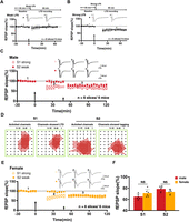
Summary of synaptic tagging of LTD in the ACC in both sexes. A, Top, A schematic diagram of the recording procedure. The summarized plot of the fEPSP slope demonstrates the induction of no-LTD for receiving weak LFS alone, which gradually increased to the baseline within 1 h. B, Top, A schematic diagram of the recording procedure. A summarized plot of the fEPSP slope demonstrates the induction of LTD for receiving strong LFS alone; strong LFS-evoked LTD lasts for at least 1 h. C, The summarized plot of the fEPSP slope shows the induction of the persistent LTD by strong LFS alone that lasts for 1.5 h of six slices/six mice (hollow circle) and demonstrates the tagged LTD induced by weak LFS if preceded, 30 min earlier, by a strong LFS and last for at least 1 h in six slices/six mice (solid circle) in the male group. Insets, Representative fEPSP traces taken at the time points indicated by numbers in the graph with calibration. Large arrows indicate the starting point of strong LFS application in S1; small arrows mark the time point of weak LFS delivery in S2. D, Left, polygonal diagrams of the channels that were activated and channels showed LTD in S1 after strong LFS. Right, polygonal diagrams of the channels that were activated and channels showed tagging in S2 after weak LFS (6 slices/6 mice). Black dots represent the 64 channels in the MED64 system and vertical lines indicate the layers in the ACC slices. The silencing responses showed after strong LFS in the S1 site and tagging LFS in the S2 site. E, The summarized plot of the fEPSP slope shows the induction of persistent LTD by strong LFS alone that lasts for 1.5 h of six slices/six mice (hollow circle) and demonstrates tagged LTD induced by weak LFS if preceded, 30 min earlier, by a strong LFS and lasts for at least 1 h in six slices/six mice (solid circle) in the female group. F, Bar histogram showing quantified data within the LTD in the S1 site and tagged LTD in the S2 site recording in the last 20 min from the male/female group. NS, no significance; error bars represent SEM.
A possible explanation for the synaptic tagging LTD phenomenon is that PRPs are generated during the process of LTD, and these PRPs are transferred between synapses. When we compared the number of active channels and LTD-showing channels after strong LFS in the S1 site or compared the number of active channels and tagged LTD-showing channels after weak LFS in the S2 site (n = 6 slices/6 mice from Fig. 2C, Fig.2D), we found that the area of channels that produced LTD due to strong LFS around the S1 were uniformly smaller than that of active channels. Interestingly, the tagging-showing channels surrounding S2 were mainly located at the superficial layer (significantly closer to the S1 site) than the original S2 active channels. This suggests that tagged LTD at the S2 site might be dependent on PRPs that were generated by and migrated from the S1 site.
To investigate whether there is any sex difference in the tagged LTD, we performed the same experiment with adult female mice. We found that tagged LTD could be induced in the ACC of female mice (S1, 70.9 ± 3.3% of the baseline at 1.5 h after strong LFS; n = 6 slices/6 mice; t(5) = 11.45; p < 0.0001; paired t test; S2, 73.0 ± 4.8% of the baseline at 1 h after weak LFS; n = 6 slices/6 mice; t(5) = 16.80; p < 0.0001; paired t test; Fig. 2E). As comparing with results from male mice, there was no significant difference in tagged LTD (S1, t(5) = 1.859; p = 0.0927; unpaired t test; S2, t(5) = 2.086; p = 0.0636; unpaired t test; Fig. 2F).
Stability and silencing responses within the ACC network in tagged LTD
During the recording of LTD, it is possible to observe silencing responses, that is, those that were active during baseline recordings showed inactive fEPSPs after LFS induction (Staubli and Ji, 1996). In the recording of tagged LTD, we found that similar to strong LFS, tagged LTD induced by weak LFS also has the appearance of silencing responses surrounding the S2 site (Fig. 3C). These silencing responses are located at the edges of the original active areas, and their amplitude of fEPSPs remained stable for 1 h during recording (Fig. 3B). Notably, LFS-induced silencing was found in only a few channels, while most of other channels remained active throughout the recording (Fig. 3A,B), ruling out the possibility that the silencing responses were caused by external instability. We counted all activated channels surrounding the S1 and S2 sites at the baseline, after strong and weak LFS (Fig. 3A, a sample slice; Fig. 3B, pool data of six slices/six mice from Fig. 2C). We found that the number of active channels surrounding S1 site decreased significantly after strong LFS. Interestingly, after weak LFS, the number of active channels at the S2 site also decreased significantly. In contrast, there was no significant change in the number of responsive channels around the S2 site after strong LFS or around the S1 site after weak LFS. This indicates that during the recording process, the LTD at both the S1 and S2 sites remained stable and the stimuli applied at the S1 and S2 sites were independent. However, the percentage of tagged LTD silencing responses (the number of silencing responses/the number of all active channels) was lower in comparison with those of strong LFS-induced LTD (Fig. 3D). A possible explanation is that although the migrated PRPs are captured and utilized, due to the limited amount of PRPs, the intensity of LTD at superficial S1 sites cannot be reached at deep S2 sites. This is consistent with the result that the depression level of tagged LTD in S2 is weaker than that of the LTD in S1 (Fig. 2C).
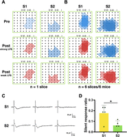
Tagged LTD showed stability, and silencing responses occurred in the S2 pathway after synaptic tagging in the ACC. A, Polygonal diagrams of the channels that were activated in the baseline state, at 30 min after strong LFS and 60 min after weak LFS of the S1 site and S2 site in one slice (blue, pre; red, post). Black dots represent the 64 channels in the MED64 system, and vertical lines indicate the layers in the ACC slice. The silent responses showed after strong LFS in the S1 site and tagging LFS in the S2 site. B, Pooled data of six slices from six mice for the spatial analysis performed as in A. Delivery of strong LFS in the S1 site resulted in a decrease in the activation area, and tagging LFS also recruited new silent responses in the ACC. C, Typical sample traces in the S1 site and S2 site at different time points (baseline, after strong LFS, after weak LFS) with calibration. D, The comparison of the number of silent responses/all channels in LTD induction between the S1 pathway and S2 pathway (5 slices/5 slices; *p = 0.0168).
The time window of synaptic tagging of LTD in the ACC
Previous studies indicate that presynaptic neuron activation triggers calcium inflow in postsynaptic neurons causing the spine to enter a temporary “tag” state of early synaptic plasticity. This state involves numerous proteins and their interactions and can last for >60 min (Redondo and Morris, 2011; Ding et al., 2021). Accordingly, we examined whether extending the interval between strong and weak stimuli affects synaptic tagging of LTD in the ACC. We modified the interval between strong LFS at S1 and weak LFS at S2 to either 60 or 90 min, with all other experimental procedures remaining consistent with those previously described (Fig. 4). We observed that synaptic tagging was still evident with a 60 min interval (S1, 55.0 ± 5.6% of the baseline at 2 h after strong LFS; n = 6 slices/6 mice; t(5) = 11.45; p < 0.0001; paired t test; S2, 61.9 ± 7.4% of the baseline at 1 h after weak LFS; n = 6 slices/6 mice; t(5) = 13.10; p < 0.0001; paired t test; Fig. 4A). When the interval was extended to 90 min, synaptic tagging of LTD was still evident in the S2 pathway (S1, 60.0 ± 4.4% of the baseline at 2.5 h after strong LFS; n = 5 slices/5 mice; t(4) = 8.483; p = 0.0011; paired t test; S2, 60.2 ± 7.3% of the baseline at 1 h after weak LFS; n = 5 slices/5 mice; t(4) = 5.591; p = 0.0050; paired t test; Fig. 4B), aligning with previous findings in the hippocampus and reports of tagged LTP in the ACC (Frey and Morris, 1997; M. G. Liu et al., 2018).

The time window of synaptic tagging of LTD. A, B, Schematic diagrams of the recording procedure for each experiment are shown at the top of the plots. Insets, Representative fEPSP traces taken at the time points indicated by numbers in the graph with calibration. Large arrows indicate the starting point of strong LFS application in S1; small arrows mark the time point of weak LFS delivery in S2. A, Pooled data showed that strong LFS at the S1 site (hollow circle) and weak LFS at the S2 site (solid circle) 1 h after strong LFS could induce LTD (6 slices/6 mice). B, The weak LFS protocol was applied to the S2 site at 90 min after the strong LFS of the S1site. Pooled data showed that weak LFS could still be affected from the strong LFS by inducing LTD (5 slices/5 mice). Strong LFS-evoked LTD lasts for 2.5 h (hollow circle), and tagged LFS-evoked LTD lasts for at least 1 h (solid circle). C, Bar histogram showing quantified data within the LTD recording at the last 20 min in the S1 site in 30, 60, and 90 min interval groups; *p = 0.0183; error bars represent SEM. D, Bar histogram showing quantified data within the tagged LTD recording in the last 20 min at the S2 site in 30, 60, and 90 min interval groups. NS, no significance; error bars represent SEM.
In addition, we compared the level of synaptic depression in the last 20 min of the S1 recording for the 30 (Fig. 2C), 60, and 90 min interval groups (S1: 30 min, 64.6 ± 2.0% of the baseline; n = 6 slices/6 mice; 60 min, 56.2 ± 3.7% of the baseline; n = 6 slices/6 mice; 90 min, 70.4 ± 3.6% of the baseline; n = 5 slices/5 mice; Fig. 4C; F(2,14) = 5.090; p = 0.0218; 30 min group vs 60 min group vs 90 min group; one-way ANOVA). Interestingly, we found that when the interval between strong and weak LFS is extended to 60 or 90 min, a slight depression can be observed at the S2 site before the weak LFS (Fig. 4A,B, data shown as solid circles). One explanation for this is that the heterosynaptic LTD around the S2 site has been induced by the strong LFS at the S1 site (Abraham et al., 2007). We found that the time this phenomenon appeared (∼40 min after strong LFS) was very similar in both the 60 min and the 90 min groups. This was not observed in the 30 min group (Fig. 2C), suggesting that the 30 min interval may not be long enough for this phenomenon.
In consideration of this finding, we used the averaged slope of fEPSP in the last 20 min before the weak LFS as the baseline (S2: 30 min, 78.7 ± 2.0% of the fEPSP slope before weak LFS; n = 6 slices/6 mice; 60 min, 71.6 ± 2.0% of the fEPSP slope before weak LFS; n = 6 slices/6 mice; 90 min, 76.4 ± 5.7% of the fEPSP slope before weak LFS; n = 5 slices/5 mice; Fig. 4D). The mean depression level of the activated channels demonstrated no difference among different intervals (F(2,14) = 1.403; p = 0.2783; 30 min group vs 60 min group vs 90 min group; one-way ANOVA). By comparing the data of S1 (Fig. 4C) and S2 (Fig. 4D) of the three groups, it is observed that the depression level of tagged LTD at the S2 site is less than that of the corresponding LTD in the S1 site (30 min, S1 vs S2, p = 0.0006; t(5) = 4.97; unpaired t test; 60 min, S1 vs S2, p = 0.0042; t(5) = 3.680; unpaired t test; 90 min, S1 vs S2, p = 0.3669; t(4) = 0.9563; unpaired t test), which is consistent with our previous suggestion that the strength of tagged LTD in the deep S2 site may not reach that of the LTD at the superficial S1 site. A more detailed analysis of the LTD-showing channels found that the percentage of the LTD-showing channels induced by the S2 pathway did not decrease at the 60 or 90 min interval group compared with that at the 30 min interval group (percentage of LTD-showing channels: 30 min group, 49 channels in 71 channels, 69.0%; 60 min group, 62 channels in total 72 channels, 86.1%; 90 min group, 59 channels in total 77 channels, 76.6%). Thus, the time window of synaptic tagging of LTD can be extended to >1 h, while the number of tagged LTD-producing channels did not decrease, which is different from that of tagged LTP (M. G. Liu et al., 2018).
mGluRs and L-voltage–gated calcium channel-dependent synaptic tagging in the ACC
The LTD in the ACC requires the involvement of relevant receptors and molecules (S. J. Kang et al., 2012; Koga et al., 2016). We wanted to know if tagged LTD also requires receptors and molecules. After the induction of LTD at the S1 site and stable recording for 20 min, that is, 10 min before the delivery of weak LFS to the S2 site, we applied antagonists of a few key molecules in glutamate neural transmission. Fifteen minutes after the weak LFS, the drugs were rinsed, and the response was continuously monitored for 1 h (Figs. 5–7). Many studies have reported that NMDA receptors play important roles in the induction of LTD (Q. Y. Chen et al., 2021; Sanderson et al., 2022). Utilizing AP-5 (50 μM), an antagonist of NMDA receptors, we found that both the synaptic tagging around the S2 site and LTD in the S1 site were intact (S1, 73.9 ± 4.8% of the baseline at 1.5 h after strong LFS; n = 5 slices/5 mice; t(4) = 4.938; p = 0.0078; paired t test; S2, 69.8 ± 8.6% of the baseline at 1 h after weak LFS; n = 5 slices/5 mice; t(4) = 5.352; p = 0.0059; paired t test; Fig. 5A). The calcium-permeable α-amino-3-hydroxy-5-methyl-4-isoxazolepropionic acid (AMPA) receptor antagonist Naspm (50 μM) affected neither the synaptic tagging around S2 nor the LTD around S1 (S1, 64.4 ± 4.4% of the baseline at 1.5 h after strong LFS; n = 5 slices/5 mice; t(4) = 6.347; p = 0.0032; paired t test; S2, 64.2 ± 10.0% of the baseline at 1 h after weak LFS; n = 5 slices/5 mice; t(4) = 15.57; p < 0.0001; paired t test; Fig. 5B). These results indicate that the tagged LTD in the ACC requires neither the NMDA receptor nor the calcium-permeable AMPA receptor. However, nimodipine (10 μM) blocked synaptic tagging of LTD around S2 without affecting the LTD in the S1 (S1, 76.2 ± 5.7% of the baseline at 1.5 h after strong LFS; n = 5 slices/5 mice; t(4) = 4.767; p = 0.0175; paired t test; S2, 101.8 ± 14.4% of the baseline at 1 h after weak LFS; n = 5 slices/5 mice; t(4) = 0.3098; p = 0.7722; paired t test; Fig. 5C), suggesting that synaptic tagging of LTD requires the involvement of L-voltage–gated calcium channels (L-VGCCs).

Synaptic tagging of LTD in the ACC depends on the activation of L-VGCCs but not the NMDA receptor. A–C, Pooled data of the fEPSP slope from all channels for both S1-strong and S2-weak inputs. We applied antagonists/inhibitors at 20 min after strong LFS of the S1 site and washout at 15 min after weak LFS to the S2 site. The large arrow indicates the starting point of strong LFS application in S1; the small arrow marks the time point of weak LFS delivery in S2. Error bars represent SEM. A, In the presence of the NMDA receptor antagonist AP-5 (50 μM), LTDs in the S1 pathway and tagging in the S2 pathway are unaffected (5 slices/5 mice). B, Naspm (50 μM), an AMPA receptor antagonist, didn't block the induction of synaptic tagging after weak LFS (5 slices/5 mice). C, The L-VGCC blocker nimodipine (10 μM) blocked the occurrence of synaptic tagging in the ACC (5 slices/5 mice). D, Bar histogram showing quantified data within the synaptic tagging recording in the last 20 min with the application of AP-5, nimodipine, and Naspm. The last 20 min of the 30 min interval group in Figure 2C was used as a control group. ***p = 0.0001; ****p < 0.0001 versus the control group; error bars represent SEM.
In addition to NMDA receptors, it has been clearly demonstrated that mGluRs play an important role in the process of LTD (Koga et al., 2016). To investigate whether the mGluRs are involved in the synaptic tagging of LTD, we first used the antagonist of mGluRs, MCPG (100 μM). We found that the synaptic tagging around the S2 site was blocked, while the LTD in the S1 site remained (S1, 86.5 ± 4.9% of the baseline at 1.5 h after strong LFS; n = 5 slices/5 mice; t(4) = 2.784; p = 0.0496; paired t test; S2, 101.7 ± 10.7% of the baseline at 1 h after weak LFS; n = 5 slices/5 mice; t(4) = 0.03822; p = 0.9713; paired t test; Fig. 6A). To further determine which subtype is involved, we used a selective antagonist of mGluR1a (LY367385, 100 μM; S1, 75.0 ± 4.4% of the baseline at 1.5 h after strong LFS; n = 5 slices/5 mice; t(4) = 11.11; p = 0.0004; paired t test; S2, 95.0 ± 8.7% of the baseline at 1 h after weak LFS; n = 5 slices/5 mice; t(4) = 1.239; p = 0.2830; paired t test; Fig. 6B), a selective antagonist of mGluR1b (CPCCOEt, 100 μM; S1, 68.4 ± 5.8% of the baseline at 1.5 h after strong LFS; n = 5 slices/5 mice; t(4) = 6.699; p = 0.0068; paired t test; S2, 96.0 ± 9.1% of the baseline at 1 h after weak LFS; n = 5 slices/5 mice; t(4) = 0.9613; p = 0.3908; paired t test; Fig. 6C), and an antagonist of mGluR5 (MPEP, 10 μM; S1, 76.4 ± 5.2% of the baseline at 1.5 h after strong LFS; n = 5 slices/5 mice; t(4) = 4.042; p = 0.0156; paired t test; S2, 70.3 ± 9.1% of the baseline at 1 h after weak LFS; n = 5 slices/5 mice; t(4) = 5.076; p = 0.0071; paired t test; Fig. 6D) and found that both mGluR1a and mGluR1b are involved in the synaptic tagging of LTD process, while mGluR5 has no such role.

mGluR1 mediate synaptic tagging of LTD in the ACC. A, MCPG (100 μM), a mGluR antagonist, blocked the induction of tagged LTD and partly affected LTD activated by strong LFS of the ACC (5 slices/5 mice). B, mGluR1a antagonist LY367385 (100 μM) blocked the occurrence of synaptic tagging-liked LTD in the ACC (5 slices/5 mice). C, CPCCOEt (100 μM), a selective mGluR1b antagonist, also significantly blocked the induction of synaptic tagging of LTD of the ACC (5 slices/5 mice). D, In the presence of the mGluR5 antagonist MPEP (10 μM), LTDs in the S1 pathway and tagging in the S2 pathway are unaffected (5 slices/5 mice). E, Bar histogram showing quantified data within the tagged LTD recording in the last 20 min with MCPG, LY367385, CPCCOEt, and MPEP. The last 20 min of the 30 min interval group in Figure 2C was used as a control group. *p = 0.0182; ****p < 0.0001 versus the control group; error bars represent SEM.
Signaling molecular-dependent synaptic tagging of LTD in the ACC
Synaptic tagging involves the interaction of multiple intracellular and extracellular signaling pathways and intracellular molecular mechanisms (Frey and Morris, 1997; Redondo et al., 2010; Sajikumar and Korte, 2011; M. G. Liu et al., 2018; P. Park et al., 2019; Zhou et al., 2023). Two major forms of LTD have been characterized in both the hippocampus and ACC: NMDA receptor-dependent LTD and mGluR-dependent LTD (mGluR-LTD; Collingridge et al., 2010; Bliss et al., 2016). Since mGluR1 plays an important role in tagged LTD of ACC, we focused on its downstream possible signaling pathways. We explored the role of some signaling molecules in synaptic tagging. The protocol we used is the same as above. We found that the protein kinase C (PKC) is critical for the induction of tagged LTD (we used an antagonist of PKC, chelerythrine chloride, 3 μM; S1, 70.8 ± 4.8% of the baseline at 1.5 h after strong LFS; n = 5 slices/5 mice; t(4) = 4.118; p = 0.0259; paired t test; S2, 95.0 ± 12.6% of the baseline at 1 h after weak LFS; n = 5 slices/5 mice; t(4) = 1.727; p = 0.1592; paired t test; Fig. 7A). Therefore, the mGluR-PKC signaling pathway is involved in the synaptic tagged LTD, which is consistent with previous reports that LTD relies on the mGluR-PKC signaling pathway (Rush et al., 2002; Jo et al., 2008). In addition, we found that the protein kinase A (PKA) is also necessary for the induction of tagged LTD, as inhibitor KT-5720 (1 μM) blocked synaptic tagging (S1, 70.8 ± 4.3% of the baseline at 1.5 h after strong LFS; n = 5 slices/5 mice; t(4) = 15.95; p = 0.0039; paired t test; S2, 98.8 ± 8.6% of the baseline at 1 h after weak LFS, n = 5 slices/5 mice; t(4) = 0.07372; p = 0.9448; paired t test; Fig. 7B). These results indicate that both PKC and PKA are required for the synaptic tagging of LTD in the ACC.

Tagged LTD in the ACC requires the activation of PKA, PKC, CaMKII, and protein phosphatase. A, Chelerythrine (5 μM), a PKC inhibitor, blocked the induction of tagged LTD after weak LFS (5 slices/5 mice). B, The PKA inhibitor KT-5720 (1 μM) blocked the occurrence of synaptic tagging in the ACC (5 slices/5 mice). C, In the presence of a CaMKII inhibitor KN-62 (10 μM), LTD in the S1 pathway and tagging in the S2 pathway are unaffected (5 slices/5 mice). D, Okadaic acid (1 μM), PP1/PP2A inhibitor, blocked the induction of synaptic tagging of LTD (5 slices/5 mice). E, Bar histogram showing quantified data within the tagged LTD recording in the last 20 min with KN-62, KT-5720, chelerythrine, and okadaic acid. The last 20 min of the 30 min interval group in Figure 2C was used as a control group. **p = 0.0014; ****p < 0.0001 versus the control group; error bars represent SEM.
Previous studies indicate that CaMKII also plays an important role in LTD (Cook et al., 2022). After perfusing reversible inhibitors of CaMKII, KN-62 (10 μM), tagged LTD of S2 gradually emerged and progressed to depression similar to S1 (S1, 69.1 ± 7.4% of the baseline at 1.5 h after strong LFS; n = 5 slices/5 mice; t(4) = 5.972; p = 0.0039; paired t test; S2, 71.6 ± 11.9% of the baseline at 1 h after weak LFS; n = 5 slices/5 mice; t(4) = 3.912; p = 0.0297; paired t test; Fig. 7C). This suggests that CaMKII is not involved in the induction of tagged LTD or the maintenance of LTD.
A previous study reported that the protein phosphatase 1 (PP1) is required for NMDA receptor-dependent LTD, while its inhibition does not block mGluR-LTD or chemical-induced LTD (Isaac, 2001). To investigate the role of PP1 in the tagged LTD, we applied okadaic acid (1 μM) before the weak LFS at S2. We found that the weak LFS failed to induce tagging and the LTD induced at S1 has also decreased (S1, 84.7 ± 5.1% of the baseline at 1.5 h after strong LFS; n = 5 slices/5 mice; t(4) = 2.945; p = 0.0422; paired t test; S2, 88.0 ± 8.1% of the baseline at 1 h after weak LFS; n = 5 slices/5 mice; t(4) = 1.616; p = 0.2474; paired t test; Fig. 7D). Therefore, PP1 is found to be necessary for the tagged LTD.
Loss of synaptic tagging of LTD after tail amputation
Considering the crucial role of ACC synapses in pain perception, we also investigated the synaptic tagging of LTD in the ACC of animal models of phantom pain. Previous studies have shown that tail amputation triggers long-term plasticity changes in the ACC and that synaptic tagging of LTP is also affected (S. J. Kang et al., 2012; M. G. Liu et al., 2018). We investigated whether synaptic tagging in the form of LTD is influenced under pathological conditions. We discovered that the synaptic tagging of LTD was completely inhibited following distal tail amputation (Fig. 8). To confirm the successful establishment of a mice model of chronic pain, we assessed the mechanical withdrawal threshold in mice before and after tail amputation. As previously reported (Zhuo, 1998; W. B. Kang et al., 2016), we found that the mechanical withdrawal threshold continued to gradually decline after the amputation (take the preoperative threshold as the baseline, paired t test; n = 5 male mice/5 female mice; Day 5: male, p = 0.6154; female, p = 0.0349; Day 8: male, p = 0.0168; female, p = 0.0195; Day 11: male, p = 0.1411; female, p = 0.0195) and remained significantly reduced at 2 weeks (Day 14: male, p = 0.0012; female, p = 0.0042; Day 17: male, p = 0.0146; female, p = 0.0075). In the slices prepared from tail amputated male mice, neither strong LFS nor tagged weak LFS elicited any significant LTD (S1, 121.7 ± 7.6% of the baseline at 1.5 h after strong LFS; n = 5 slices/5 mice; t(4) = 5.627; p = 0.0049; paired t test; S2, 102.7 ± 9.7% of the baseline at 1 h after weak LFS; n = 5 slices/5 mice; t(4) = 0.6194; p = 0.5692; paired t test; Fig. 8B). The activated channels were divided into channels with LTD (S1, one channel; induction rate, 1.7%; S2, four channels; induction rate, 6.3%) or no-LTD channels (S1, 59 channels; induction rate, 98.3%; S2, 60 channels; induction rate, 93.7%; Fig. 8C,D).

Tail amputation blocked synaptic tagging of the ACC in both sexes. A, A schematic diagram of a tail amputation model. B, A schematic diagram of the recording procedure for the experiment is shown at the top of the plot. Insets, Representative fEPSP traces taken at the time points indicated by numbers in the graph with calibration. Pooled data of the fEPSP slope from all channels for both S1-strong and S2-weak inputs. After strong stimulation delivery and recording for 30 min, weak stimulation was given and then recording for 1 h. The large arrow indicates the starting point of strong LFS application in S1; the small arrow marks the time point of weak LFS delivery in S2. The induction of persistent no-LTD by strong LFS or no synaptic tagged LTD by weak LFS in male mice after amputation (5 slices/5 mice). C, D, Individual data presentation of no-LTD recording in the S1 site and no synaptic tagging recording in the S2 site in the ACC. Among five slices analyzed, 58 showed no-LTD, and 60 channels showed no synaptic tagged LTD. E, Pooled data of the fEPSP slope from all channels for both S1-strong and S2-weak inputs. After strong stimulation delivery and recording for 30 min, weak stimulation was given, and then recording was continued for 1 h. The induction of persistent no-LTD by strong LFS or no synaptic tagged LTD by weak LFS in female mice after amputation (5 slices/5 mice). F, Bar histogram showing quantified data of the ratio of no-LTD in the S1 site and no-tagged LTD in the S2 site recording from the male/female group. NS, no significance; error bars represent SEM. G, H, Individual data presentation of no-LTD recording in the S1 site and no synaptic tagging recording in the S2 site in the ACC of female mice. Among five slices analyzed, 63 showed no-LTD, and 98 channels showed no synaptic tagged LTD.
We aimed to determine if the changes in synaptic plasticity caused by tail amputation vary between sexes. We replicated the model in female mice (S1, 93.2 ± 3.4% of the baseline at 1.5 h after strong LFS; n = 5 slices/5 mice; t(4) = 3.171; p = 0.0338; paired t test; S2, 102.5 ± 7.1% of the baseline at 1 h after weak LFS; n = 5 slices/5 mice; t(4) = 0.1066; p = 0.9202; paired t test; Fig. 8E). We also quantified the channels with LTD (S1, 10 channels; induction rate, 13.7%; S2, 26 channels; induction rate, 21.0%) and without LTD (S1, 63 channels; induction rate, 86.3%; S2, 98 channels; induction rate, 79.0%) in the female group (Fig. 8G,H). There was no significant difference in the induction rate of LTD/tagged LTD between male and female mice models of tail amputation (S1, t(4) = 1.292; p = 0.2324; S2, t(4) = 0.5265; p = 0.6128; Fig. 8F). Additionally, we observed no silencing response during this process. We conclude that both the weak LFS-induced tagged LTD and the strong LFS-induced LTD are occluded by peripheral nerve injury. This finding provides new theoretical insights into the neural mechanisms of chronic pain at the synaptic level.
Discussion
Phantom pain and amputation-related brain disorders remain significant medical challenges. In this study, we show that the ACC, a key cortical region for pain and emotion, is capable of undergoing tagged LTD, and this form of tagged LTD is reduced following tail amputation. To our knowledge, this is the first report of synaptic tagging of LTD at ACC synapses in adult animals. Interestingly, previous research has identified synaptic tagging of LTP, a key form of heterosynaptic LTP in the hippocampus, at ACC synapses, which is also impacted by tail amputation (M. G. Liu et al., 2018). These results suggest that excitatory synapses in the ACC can exhibit biphasic synaptic tagging. Homosynaptic LTD has previously been studied in this region following amputation (S. J. Kang et al., 2012). Combined with this earlier study, it appears that both homosynaptic and heterosynaptic LTD are significantly affected in the ACC postamputation. The reduction of these different forms of LTD could lead to increased cortical excitation in the adult cortex following amputation (Zhuo, 2008, 2016b). Our findings further provide new insights into the synaptic mechanisms at cortical synapses that may underlie pathological changes in amputees.
Synaptic tagging in the hippocampus
The hippocampus has long been recognized as a pivotal area for studying functional neuroplasticity, hosting various models of synaptic plasticity such as LTP and LTD. These processes play diverse cognitive roles in reinforcement, relearning, and memory association (see Introduction). Both LTP and LTD can manifest in transient early forms (early-LTP/LTD) or persistent late forms (late-LTP/LTD). They are initiated by different induction protocols, and their mechanisms vary, particularly in terms of protein synthesis requirements and the involvement of key molecules (Y. Y. Huang et al., 1994; Y. Y. Huang and Kandel, 1996; T. Chen et al., 2014). Synaptic tagging, first identified in the hippocampus, involves a weak tetanus creating a “tag” that captures newly synthesized PRPs triggered by a strong tetanus (Reymann and Frey, 2007; Redondo and Morris, 2011; Vecsey et al., 2018). Several candidate proteins have been proposed, as they are involved in the maintenance of tagging in the hippocampus, such as CaMKII, β-actin, calcium-permeable AMPA receptors, and PKA. In the present study, we observed that synapses in the ACC can also undergo tagging of LTD. These findings suggest that synaptic tagging can be biphasic. To our knowledge, reports of synaptic tagging of LTD specifically in the hippocampus are still limited.
Tagged LTD: a heterosynaptic LTD
Recently, the Bito group discovered inverse synaptic tagging of Arc/Arg3.1 via CaMKIIβ (Okuno et al., 2012). Arc/Arg3.1, as an immediate early gene product, can be captured and preferentially targeted to mark nonpotentiated synapses, which prevents these synapses from strengthening. It is possible that this mechanism works together with synaptic tagging to regulate the proportion of active synapses (Okuno et al., 2018). The “tag” presented by synapses expressing early-LTD may also capture protein synthesis products produced during late-LTD induction in a time window, in other words, tagged LTD is also present (Sajikumar and Frey, 2004; Bin Ibrahim et al., 2022). Sajikumar et al. (2005) reported a phenomenon known as “cross tagging,” where late-LTP in one input can transform early-phase LTD into late-phase LTD in a separate input and vice versa. Unlike heterosynaptic LTP, synaptic tagging of LTD has been less explored. In our study, we demonstrate for the first time that LTD in the ACC can also undergo synaptic tagging. This confirms the existence of bidirectional synaptic tagging in the ACC. We found that weak repetitive stimulation cannot induce long-lasting synaptic depression (at least 20% less than the baseline) as standard LFS at 1 Hz for 15 min does. However, a weak stimulation can induce LTD if it follows a heterosynaptic LTD. Similar to tagged LTP, tagged LTD can also be produced and sustained for at least 1 h due to the presence of PRPs and other associated molecules. This process also involves the emergence of silencing responses. We also confirmed that tagged LTD is similarly present in ACC of female mice. However, the specific differences between sexes need to be further studied (R. H. Liu et al., 2020).
Different forms of synaptic depression in the adult ACC
Using different recording methods (whole-cell patch-clamp and field recording), previous studies demonstrated that ACC synapses can undergo different forms of LTD (see Introduction). LFS in the field recording showed that 1–5 Hz repetitive stimulation can induce LTD of synaptic responses in the ACC (Wei et al., 1999; S. J. Kang et al., 2012; Xue et al., 2021). This form of LTD is sensitive to L-VGCCs, mGluRs, and NMDA receptors (Wei et al., 1999; Xue et al., 2021). In contrast, LTD induced by paired-induction protocol in the whole-cell patch recording is mainly NMDA receptor-dependent (Toyoda et al., 2005, 2007), and the expression of LTD required the involvement of AMPA subtype GluA2 receptors (Toyoda et al., 2005, 2007). In the present study, we found that tagged LTD in the field recording system is sensitive to inhibition of both L-VGCCs and mGluRs, while inhibition of NMDA receptors did not affect the induction. These findings suggest that tagged LTD and LTD do not share the same synaptic mechanisms.
Previous studies demonstrated that tagged LTP in the hippocampus is independent of the activation of NMDA receptors (P. Park et al., 2019) but requires Group Ⅲ mGluRs (GluR4 and 7; Dasgupta et al., 2020). This is similar to the tagged LTD since the inhibition of the NMDA receptors failed to induce changes in the tagged LTD, while the inhibition of mGluRs affected the tagged LTD. However, few studies investigate the role of L-VGCCs in the synaptic tagging of LTP.
mGluRs and intracellular signaling pathway in synaptic depression
mGluR-LTD is a form of synaptic plasticity needed for learning and memory, which requires L-VGCCs (Chiamulera et al., 2021; Q. Y. Chen et al., 2021). In the hippocampal CA1 and CA2 regions, the selective mGluR agonist (RS)-3,5-dihydroxyphenylglycine (DHPG) induces LTD, and this mGluR-LTD can be expressed by changes in the number of postsynaptic AMPA receptors or reduced probability of neurotransmitter release (Sanderson et al., 2022; Samadi et al., 2023). In the insular cortex (IC) of adult mice, tail amputation leads to the loss of LFS-induced LTD without affecting DHPG-induced LTD (M. G. Liu and Zhuo, 2014). In the ACC, we found that DHPG could induce LTD (S. J. Kang et al., 2012). Fragile X mental retardation protein (FMRP) plays an important role in the regulation of synaptic protein synthesis, and its deficiency can lead to learning and behavior disorders (Bear et al., 2004; Koga et al., 2015a). In the ACC neurons, activation of Group Ⅰ mGluRs may upregulate FMRP through the CREB signaling pathway (Wang et al., 2012). Our results prove that mGluRs, especially mGluR1, play an important role in tagged LTD of ACC. In addition, PKA is particularly important for synaptic tagging, and blocking PKA anchoring impairs synaptic tagging in the hippocampus (T. Huang et al., 2006; A. J. Park et al., 2014). CaMKII is important for long-term plasticity and is required by both NMDA receptor-dependent LTD and mGluR-LTD (Cook et al., 2022). The PKC pathway in mGluR-LTD has additionally been extensively reported, with our experimental results in confirmation with these dynamics.
PP1 plays a key role in NMDA receptor-dependent LTD in the hippocampus (Lisman, 1989; Mulkey et al., 1994). Interestingly, chemical-induced hippocampal LTD by applying DHPG is PP1-independent (Isaac, 2001; Morishita et al., 2001). In the present study, we reproduced previous findings that field recording of LTD in the ACC is mGluR-dependent (Koga et al., 2016). This form of LTD is not blocked by AP-5 (S. J. Kang et al., 2012). In the present study, we found that PP1 is required for mGluR-dependent tagged LTD in the ACC. This new finding suggests that PP1 plays critical roles in different forms of LTDs in the hippocampus and ACC. Interestingly, a recent study by Chang et al. (2015) using whole-cell patch-clamp found that NMDA receptor-dependent LTD in the ACC was blocked by PP1 inhibitor okadaic acid.
Functional implications of synaptic tagging of LTD
Cumulative evidence demonstrates that ACC and IC show enhanced excitatory synaptic transmission and neuronal excitability after peripheral injury and amputation which contribute to the induction and maintenance of persistent pain and are associated with an enhancement of long-term behavioral responses (Bliss et al., 2016; Zhuo, 2016a). This cortical excitation shares some common synaptic mechanisms with different forms of LTP studies in vitro. It has been suggested that presynaptic LTP in the ACC and IC contribute to behavioral anxiety/depression caused by injury, with postsynaptic LTP more likely to contribute to behavioral sensitization, allodynia, and hyperalgesia (Koga et al., 2015b; Bliss et al., 2016). Consistent with this discovery of LTP, the loss of LTD has been consistently found in these areas after peripheral injury and amputation.
Under normal physiological conditions, LTD provides an important mechanism to “reset” these enhanced synapses, ensuring that cortical synapses and circuits are maintained at the baseline level allowing for the processing of multiple activities. After peripheral injury and/or amputation, abnormal neuronal activity from the periphery triggers long-term transcriptional and translational changes in ACC synapses. This may be due to two mechanisms: enhanced synaptic transmission and the loss of the ability to undergo LTD. These mechanisms may work together on synaptic enhancement. In addition to changes in LTP/LTD at the same synapses (homosynaptic form of plasticity), our recent and present results with tagged experiments indicate that adjacent synapses (heterosynaptic form) also undergo similar changes. This new finding provides a possible synaptic mechanism for the network changes within the ACC after amputation. The changes in synaptic plasticity not only happen among primary synapses that receive direct projections from peripheral inputs but also in adjacent, heterosynaptic connections.
It is well known that amputation leads to a cortical reorganization associated with phantom pain and sensation (M. M. Merzenich et al., 1984; Kaas, 1991; Pons et al., 1991; M. Merzenich, 1998; Montoya et al., 1998; Kaas et al., 1999). Since most of these cortical reorganizations are found months or years after amputation, it is likely that synaptic changes reported here occur before the structural reorganization in the cortex. A recent study has reported that motor learning induced new dendritic spines in the motor cortex of adult mice at 8–24 h after training (Qiao et al., 2022). We cannot rule out the possibility that similar changes in synaptic spines may occur before the loss of synaptic tagging of LTD or at the same period. Future studies are clearly needed to address this question.
The current findings suggest that the reduction of synaptic tagging of LTD could provide a mechanism for the spread of cortical excitation across heterosynaptic sites. These interactions may occur presynaptically and postsynaptically, potentially contributing to chronic pain and associated anxiety/depression, which are commonly reported among amputees (McKechnie and John, 2014; Raichle et al., 2015; Bennett, 2016). Further studies are necessary to elucidate the precise molecular and cellular mechanisms behind the loss of synaptic tagging postamputation, as well as its impact on behavioral changes.
Footnotes
- Received January 5, 2024.
- Revision received July 2, 2024.
- Accepted July 5, 2024.
-
We thank the EJLB-CIHR Michael Smith Chair in Neurosciences and Mental Health in Canada, Canada Research Chair, Ontario-China Research and Innovation Fund (OCRIF), and Canadian Institute for Health Research operating and project grants (MOP-124807, PJT-148648, and 419286) for funding support to M.Z. X-H.L. was supported by grants from Fundamental Research Funds for the Central Universities (xxj032022013 and xzy012022046). We are grateful to Emily England and Danielle Zhuo for helping review the manuscript and providing valuable suggestions.
-
The authors declare no competing financial interests.
- Correspondence should be addressed to Min Zhuo at min.zhuo{at}utoronto.ca.
- Copyright © 2024 the authors




