The Lysine Acetyltransferase PCAF Functionally Interacts with Estrogen Receptor Alpha in the Hippocampus of Gonadally Intact Male—But Not Female—Rats to Enhance Short-Term Memory
- Krista A. Mitchnick1,2,
- Kate Nicholson2,3,
- Cassidy Wideman1,2,
- Kristen Jardine1,2,
- Rhiannon Jamieson-Williams1,
- Samantha D. Creighton1,2,
- Allison Lacoursiere1,
- Ciro Milite4,
- Sabrina Castellano4,
- Gianluca Sbardella4,
- Neil J. MacLusky2,3,
- Elena Choleris1,2, and
- Boyer D. Winters1,2
- 1Department of Psychology, University of Guelph, Guelph, Ontario N1G 2W1, Canada
- 2Collaborative Neuroscience Program, University of Guelph, Guelph, Ontario N1G 2W1, Canada
- 3Department of Biomedical Sciences, University of Guelph, Guelph, Ontario N1G 2W1, Canada
- 4Department of Pharmacy, Epigenetic Med Chem Lab, University of Salerno, Fisciano I-84084, Italy
-
Author contributions: K.A.M., K.N., N.J.M., E.C., and B.D.W. designed research; K.A.M., K.N., C.W., K.J., R.J.-W., S.D.C., and A.L. performed research; C.M., S.C., and G.S. contributed unpublished reagents/analytic tools; K.A.M., K.N., and C.W. analyzed data; K.A.M. wrote the paper.
Abstract
Acetylation of histone proteins by histone acetyltransferases (HATs), and the resultant change in gene expression, is a well-established mechanism necessary for long-term memory (LTM) consolidation, which is not required for short-term memory (STM). However, we previously demonstrated that the HAT p300/CBP-associated factor (PCAF) also influences hippocampus (HPC)-dependent STM in male rats. In addition to their epigenetic activity, HATs acetylate nonhistone proteins involved in nongenomic cellular processes, such as estrogen receptors (ERs). Given that ERs have rapid, nongenomic effects on HPC-dependent STM, we investigated the potential interaction between ERs and PCAF for STM mediated by the dorsal hippocampus (dHPC). Using a series of pharmacological agents administered directly into the dHPC, we reveal a functional interaction between PCAF and ERα in the facilitation of short-term object-in-place memory in male but not female rats. This interaction was specific to ERα, while ERβ agonism did not enhance STM. It was further specific to dHPC STM, as the effect was not present in the dHPC for LTM or in the perirhinal cortex. Further, while STM required local (i.e., dHPC) estrogen synthesis, the facilitatory interaction effect appeared independent of estrogens. Finally, western blot analyses demonstrated that PCAF activation in the dHPC rapidly (5 min) activated downstream estrogen-related cell signaling kinases (c-Jun N-terminal kinase and extracellular signal-related kinase). Collectively, these findings indicate that PCAF, which is typically implicated in LTM through epigenetic processes, also influences STM in the dHPC, possibly via nongenomic ER activity. Critically, this novel PCAF–ER interaction might exist as a male-specific mechanism supporting STM.
Significance Statement
Due to their ability to modulate gene expression, epigenetic mechanisms are necessary for long-term, but not short-term, memory. Recently, the histone acetyltransferase PCAF has shown necessity in the hippocampus (HPC) for short-term object memory, but its mechanism of action is unknown. Here, we demonstrated a novel functional interaction between PCAF and estrogen receptors (ERs) in the HPC of gonadally intact male—but not female—rats that enhanced short-term object memory. Furthermore, enhancing PCAF led to rapid activation of downstream ER-related signaling cascades, suggesting that PCAF might activate ERs similar to the effects of estrogens. Indeed, the PCAF-ER interaction appeared estrogen-independent. These results reveal a novel mechanism mediating short-term memory enhancement that appears to be specific to males.
Introduction
Extensive evidence demonstrates the requirement of histone acetylation for long-term memory (LTM). Histone acetyltransferases (HATs) catalyze the addition of acetyl groups to various lysine residues on histone tails, typically promoting gene expression by making the DNA accessible to transcriptional machinery or by recruiting other factors (Peixoto and Abel, 2013). Interestingly, our group (Mitchnick et al., 2016) and others (Maurice et al., 2008) have demonstrated the necessity of one specific HAT, PCAF (p300/CBP-associated factor), in short-term memory (STM) as well. Specifically, male and female PCAF knock-out mice display short-term (3 min) object-in-place (OiP) memory deficits (Maurice et al., 2008). Similarly, male rats administered the small-molecule PCAF inhibitor embelin directly into the dorsal (d) hippocampus (HPC), but not the perirhinal cortex (PRh), also exhibiting short-term (5 min) OiP memory impairments (Mitchnick et al., 2016). Furthermore, pharmacological activation of PCAF has been recently demonstrated to enhance short-term (5 min) object location memory in young male and female 3xTG Alzheimer's disease mice (Creighton et al., 2022). Because STM does not require de novo gene expression (Davis and Squire, 1984), it is unlikely that PCAF is acting at the genomic level in these instances (Mitchnick et al., 2016).
Although often highlighted for their role in acetylating histone proteins, HATs also acetylate lysine residues on a plethora of nonhistone proteins and are more broadly referred to as lysine acetyltransferases (KATs; You et al., 2012; Downey, 2021). For example, PCAF has been shown to bind, in a ligand-dependent manner, to the DNA-binding domain of several nuclear receptors, including estrogen receptor alpha (ERα), exhibiting its role as a nuclear receptor coactivator (Blanco et al., 1998). Similarly, PCAF has been reported to acetylate the orphan nuclear receptor estrogen-related-receptor alpha (ERRα) on several sites of the DNA-binding domain (Wilson et al., 2010).
Estrogens and ERs, including the classic nuclear ERs (ERα, ER β) and the G-protein–coupled ER (GPER), are widely implicated in LTM (≥24 h), a process known to require gene expression (Fernandez et al., 2008; Liu et al., 2008; Hammond et al., 2009; Fortress et al., 2013; Pereira et al., 2014; Kim et al., 2016; Mitchnick et al., 2019; de Souza et al., 2021). Several groups have also demonstrated that 17β-estradiol and ER activity can cause rapid effects in male and female rodents, including modulating downstream signaling cascades, such as extracellular signal-related kinase (ERK) and c-Jun-terminal kinase (JNK), synaptic structures (e.g., dendritic spines), and ultimately enhancing STM (≤4 h; Walf et al., 2006; Frye et al., 2007; Clipperton et al., 2008; Jacome et al., 2010; Phan et al., 2012, 2015; Kim et al., 2016, 2019; Lymer et al., 2017; Frick et al., 2018; Sheppard et al., 2018, 2021, 2023; Mitchnick et al., 2019; Kuun-Hadden et al., 2023). Although activational effects of PCAF on both ERα and ERRα have been shown to modulate gene transcription (Blanco et al., 1998; Wilson et al., 2010), its action on nuclear hormone receptors suggests that PCAF could also stimulate cytoplasmic or membrane-bound ERs, causing rapid, nongenomic effects, such as the activation of cell signaling kinases, to support STM.
To test this hypothesis, we investigated a potential interplay between PCAF and ERs in short-term OiP memory in gonadally intact male and female rats. In males, behavioral experiments using intra-dHPC administration of compounds to enhance or inhibit the function of PCAF, ERα, and ERβ indicated that the modulation of hippocampal STM by PCAF is mediated through functional interactions with ERα. Evidence for this interaction was not found in PRh. Furthermore, we found dHPC estrogen biosynthesis to be necessary for STM, but not for the functional interaction between PCAF and ERα. Finally, Western blot analyses suggested that PCAF activation enhances P-JNK and P-ERK levels. Surprisingly, in females, PCAF activation within the dHPC did not enhance STM. Collectively, our data indicate that the lysine acetyltransferase PCAF functionally interacts with and can have activational effects on ERα in the male rat dHPC, in a potentially estrogen-independent manner, to support STM. These mnemonic effects may be mediated by PCAF activation-induced increases in JNK and/or ERK signaling. Critically, these effects are sex-specific.
Materials and Methods
Subjects
Adult male and female Long–Evans rats (Charles River Laboratories), weighing 270–300 g (∼3 months) at the start of testing, were used for all experiments. Experiments were conducted and analyzed separately for males and females. Rats were pair housed (within sex) and maintained on a 12 h reverse light/dark cycle (08:00 h lights off; 20:00 h lights on), and behavioral testing occurred during the rats’ waking hours (dark phase). They were maintained at an 85–90% free-feeding body weight and fed after testing was completed on experiment days. Water was available ad libitum. All procedures adhered to the guidelines of the Canadian Council on Animal Care and were approved by the Animal Care Committee at the University of Guelph.
Surgical procedures
All rats underwent intracranial surgical cannula implantation targeting the dHPC (Mitchnick et al., 2016). One additional set of males underwent intracranial surgical cannula implantation targeting PRh. Prior to surgery, rats were anesthetized with isoflurane (Benson Medical Industries) and received a systemic subcutaneous injection of the analgesic meloxicam (5 mg/ml; Boehringer Ingelheim). Rats were secured in a stereotaxic frame with the incisor bar placed at −3.3 mm (Kopf Instruments). The scalp was incised and retracted to expose the skull. Holes were drilled into the skull, and 22 gauge indwelling cannula guides were implanted according to the following coordinates relative to the skull at the bregma (Paxinos and Watson, 1998): anteroposterior, −3.8 mm; lateral, ±2.5 mm; and dorsoventral, −2.5 mm for dHPC placements and anteroposterior, −5.5 mm; lateral, ±6.6 mm; and dorsoventral, −7.0 mm for PRh placements. The guide cannulas were anchored to the skull by four jeweler screws and dental acrylic. Dummy cannulas (0.36 mm diameter) were placed into the guide cannulas where they remained, except during infusions. Dummy cannulas were cut to extend 1.1 mm past the end of the guide cannula. The scalp was sutured postsurgery. Rats rested for 7–10 d to recover before testing.
OiP procedure
The behavioral procedures and apparatus have been described previously (Mitchnick et al., 2015, 2016). Figure 1 depicts the OiP task with a representative object set; four objects varying in size (7–20 cm), color, material (glass, aluminum, meal, ceramic, and plastic), and texture were placed in each corner of the open field, 5 cm away from the walls. The objects were washed with 50% ethanol between trials. The open field (60 × 60 × 36 cm3) was constructed from white, plastic-coated corrugated cardboard. The apparatus walls and floors were bare. Spatial cues were present in the testing room (i.e., party decorations on the walls, shelving, colored door), and a ceiling-mounted white light illuminated the room. All rats experienced two 5 min habituation sessions on successive days, allowing them to explore the empty open field, followed by a mock microinfusion (i.e., pump on but not connected to indwelling infusers/tubing).

OiP parameters. Schematic of the OiP task and running parameters with a representative object set. Two objects are switched during the choice phase (right side in the image), which control rats will preferentially explore. With a 5 min sample phase, rats successfully remember 24 h later (Normal Learning Conditions). This allows the assessment of memory-impairing agents. When the sample phase is reduced to 3 and 2 min, rats do not remember 24 h and 20 min later, respectively, allowing for the assessment of memory-enhancing agents. Figure reproduced from Mitchnick et al. (2016).
Depending on the experiment, rats received one or two dHPC microinfusions, before or after the sample phase (presample; postsample; Table 1). For experiments assessing the impairing effects of an agent, the sample phase was 5 min in duration. During the sample phase, rats were allowed to freely explore four different novel objects (Mitchnick et al., 2016; Fig. 1). To assess LTM, rats experienced a 2 min choice phase 24 h later, where they viewed the same four objects, but two objects had switched locations (right or left, counterbalanced) creating a “novel side” (Fig. 1a). Preference for the objects on the novel side was interpreted as being indicative of memory. To assess STM, the retention delay between the sample and choice phases was 20 min. The assessment of memory enhancement was accomplished with a suboptimal sample phase (i.e., reducing the sample phase duration such that control rats do not remember following a retention delay). Specifically, to assess LTM enhancing effects (24 h delay), a 3 min sample phase was used, whereas a 2 min sample phase was used to assess STM (20 min delay) enhancements (Fig. 1).
Inhibitor and activator compounds and infusion parameters
Due to the within-subject design of our experiments, an experiment assessing the effects of one pharmacological agent had two trials (i.e., all rats received a control infusion and a drug infusion, counterbalanced). Experiments assessing the interaction of two pharmacological agents had four trials (i.e., control/control, Drug 1/control, control/Drug 2, Drug 1/Drug 2). Trials were separated by a minimum of 2 d to reduce interference from previous object sets, as has been done previously with this paradigm (Mitchnick et al., 2015, 2016, 2019).
Inhibitors/activators and infusion parameters
All inhibitors, activators, and control compounds are listed in Table 1, including their mechanisms of action, dose, infusion parameters, and references. Compounds were dissolved in their respective control solutions. Infusions were delivered by a Harvard Apparatus precision syringe pump set to administer infusions at a rate of 0.5 μl/min. Two 1 μl Hamilton syringes were secured to the pump, and propylene tubing holding infusion cannulas were attached. Infusions lasted 2 min (total of 1 μl of infusate/hemisphere). The infusion cannulas were left in for an additional 1.5 min to allow for the diffusion of the infusate, and dummy cannulas were returned following each infusion. All infusions were performed in the dark under red light.
Wherever possible, compounds were delivered immediately after the sample phase to restrict their activity to the memory consolidation period. However, for interaction experiments where multiple compounds were utilized, one compound was administered postsample, while the other was administered presample to reduce possible tissue damage from successive infusions. PCAF-modulating compounds were always administered prior to ERs/estrogen synthesis-modulating compounds, for consistency. PCAF-modulating compounds had different presample administration parameters to accommodate their various properties. Specifically, embelin's effects on memory have been found to occur rapidly (Mitchnick et al., 2016), and therefore it was administered immediately presample. Letrozole was administered 20 min presample according to previously published parameters in rats (Sato and Woolley, 2016; Mitchnick et al., 2019). SPV106 was administered 3 d presample according to previously published parameters in mice (Wei et al., 2012; Creighton et al., 2022) and our pilot data in rats where no memory-enhancing effects were found if administered immediately postsample.
A minimum of 2 d between trials was sufficient for elimination of several compounds, including PPT and DPN which are eliminated in ∼25 h (when administered to rats subcutaneously; Sepehr et al., 2012). The intertrial interval was longer for certain experiments due to the mode of action and half-life of other compounds; specifically, 5 d were left between infusions of SPV106 as infusions were given 3 d presample, 7 d were left between infusions of ICI-182-780 to allow ERs to repopulate (Long and Nephew, 2006), and 13 d were left between letrozole infusions due to its longer half-life (Mitwally and Casper, 2001) and our pilot experiments (Table 1).
OiP data analysis
The sample and choice phases were recorded by a ceiling-mounted camera, and exploration, scored by an experimenter blind to the treatment conditions, was defined as directing the nose to the objects at a distance of <2 cm and/or touching it with the nose. Novelty preference was used as an indicator of memory and was calculated by the following discrimination ratio (DR): exploration of displaced objects minus exploration of familiar-sided objects divided by total object exploration. Although the total 2 min of choice exploration was recorded and scored, DRs were calculated from the exploration levels in the first minute only, during which object discrimination is typically most robust (Dix and Aggleton, 1999). For experiments assessing two drug conditions and two delay periods, a 2 × 2 repeated-measure analysis of variance (ANOVA) was employed, with DR as the dependent measure. Dictated by our hypotheses, specific planned comparisons (paired t tests) were conducted. For experiments that assessed the effects of a compound across different retention delays, two comparisons were planned: drug versus vehicle STM and drug versus vehicle LTM. For experiments assessing the combined effect of two compounds, three comparisons were planned: vehicle/inhibiting compound versus vehicle, enhancing compound/vehicle versus vehicle/vehicle, and enhancing compound/vehicle versus enhancing compound/inhibiting compound. For dose response experiments (one-way ANOVA), each dose condition was compared with the vehicle condition. The only exception was the assessment of a single compound on only one retention delay, where only a paired-sample t test was conducted between conditions. Additionally, total sample and choice object exploration levels for both drug and control conditions were examined in each testing phase to assess potential preexisting or drug-induced nonmnemonic differences; specifically, paired-sample t tests were run comparing the total sample or choice exploration for control and drug conditions. Control analyses were nonsignificant unless otherwise stated. Descriptive statistics can be found in Table 2. All analyses were conducted in SPSS 27.0 as two-tailed tests with a confidence interval of 0.05. False discovery rate (FDR) correction (Benjamini–Hochberg algorithm) was applied to each experiment/analysis with multiple comparisons, and corrected p values are reported. The FDR was chosen as it strikes a balance between Type 1 and Type 2 errors, compared with very stringent methods for multiple-comparison corrections (e.g., Bonferroni’s; Menyhart et al., 2021).
Behavioral exploration amounts
Histology
Brains from rats used in behavioral experiments were collected after behavioral testing and processed as previously described (Mitchnick et al., 2015, 2016) to confirm cannula placements. Specifically, rats were anesthetized by an intraperitoneal injection of 2 ml of Euthansol (340 mg/ml; Schering Canada) and perfused transcardially with phosphate-buffered saline (PBS), pH7.4, followed by 4% formalin, pH7.4 (EMD). The brains were then removed, postfixed in 4% formalin at 4°C (minimum of 1 week), and then immersed in 20% sucrose in PBS until they sank (minimum of 10 h). The brains were sectioned (55 μm) on a cryostat, through the extent of HPC or PRh, and every third section was mounted on a gelatin-coated glass slide and stained with thionin. Verification of cannula placements was carried out using light microscopy (1.25× magnification).
Protein isolation and Western blot procedures
Twenty-one adult male Long–Evans rats (Charles River) received intra-dHPC surgical cannula implantation (as described above). Following recovery and habituation to the apparatus (as described above), rats were assigned to one of the three groups (n = 7/group). The control (VEH-VEH) group received a vehicle microinfusion (1% DMSO) 3 d prior to the OiP sample phase and a second vehicle infusion (10% DMSO) immediately preceding the sample phase. The first experimental group (SPV-VEH) group received a microinfusion of SPV106 3 d presample and a vehicle microinfusion (10% DMSO) immediately presample. The second experimental group (SPV-ICI) received a 3 d presample dHPC microinfusion of SPV106 (0.4 μg/μl) and an immediate presample dHPC microinfusion of ICI-182-780 at a low dose (0.01 μg/μl) that is behaviorally ineffective in the OiP task. The drug doses and infusion parameters paralleled the behavioral experiments (Table 1), with the exception of ICI-182-780 given immediately presample rather than immediately postsample to accommodate the shortened 5 min delay. Five minutes following the sample phase, all rats were briefly exposed to CO2 and their brains quickly removed. The entire, bilateral dHPC was excised, placed in a tube, and immediately frozen on dry ice (Mitchnick et al., 2015, 2016, 2019). The tissue was maintained at −80°C until further processing. Tissue preparation procedures have been described previously (Mitchnick et al., 2019). Tissue samples were homogenized in protein lysis buffer (1% Triton X-100, 50 mM Tris, 150 mM sodium chloride), pH 7.5, with dissolved Pierce Protease Inhibitor EDTA-free mini tablets (Thermo Fisher Scientific) and Na3VO4 (5 μM). The homogenates were placed on ice, briefly sonicated, given 20 min to rock on ice for protein isolation, and centrifuged two times at 17,530 × g for 15 min at 4°C. Protein concentrations were determined using a Bradford assay (Bradford, 1976), and lysates were stored in small aliquots at −80°C until Western blotting was performed. The protocol for Western blotting has been described previously (Mendell et al., 2016; Mitchnick et al., 2019). Briefly, 30 μg of total protein for each sample was loaded onto 10% sodium dodecyl sulfate polyacrylamide gels, and electrophoresis was performed using Mini-PROTEAN Tetra cells (Bio-Rad Laboratories). The Trans-Blot SD Turbo transfer apparatus (Bio-Rad Laboratories) was used to transfer proteins onto 0.45 μM nitrocellulose (Bio-Rad Laboratories), with transfer conditions of 30 min at 25 V (constant voltage). Following the transfer, membranes were briefly rinsed in tris-buffered saline with 0.1% Tween-20 (TBS-T) and blocked in either 5% nonfat milk, 5% bovine serum albumin (BSA; Thermo Fisher Scientific) or a combination of milk and BSA for 1.5 h. Membranes were rinsed twice for 5 min in TBS-T and incubated on an orbital shaker overnight at 4°C in primary antibody for phospho-ERK1/2 [1:2,000; Cell Signaling Technologies (CST); 9101], ERK1/2 (1:2,000; CST; 9102S), phospho-JNK (1:2,000, CST; 4668S), JNK (1:400 Santa Cruz Biotechnology; sc-7345), or beta-actin (1:5,000; CST; 3700S). Blots were rinsed twice for 5 min in TBS-T, incubated for 1 h at room temperature with horseradish peroxidase (HRP)-conjugated goat anti-mouse (1:2,500; CST; 7076) or goat anti-rabbit IgG (1:2,500; CST; #074) in milk or BSA, and then rinsed twice for 5 min and twice for 10 min in TBS-T. Proteins of interest were visualized with Luminata Forte HRP substrate (Millipore Canada) on a ChemiDoc MP Imaging System (Bio-Rad Laboratories). Densitometry was performed using the Image Lab v6.1 software (Bio-Rad Laboratories).
Western blot data analysis
A 2 × 3 ANOVA (isoform × group) was performed for Western blot densitometric quantitative analysis. Data were collapsed across isoforms when no main effect of isoform was present. Based on our hypotheses, two comparisons were planned (independent t tests): SPV106/vehicle versus vehicle/vehicle and SPV106/vehicle versus SPV106/ICI-182-780. Statistical significance was set to p < 0.05. FDR (Benjamini–Hochberg algorithm) was applied to account for multiple comparisons, and the corrected p value is reported.
Results
Histology
The infusion needle tips in rats with bilateral dHPC cannulas terminated in the dHPC and were consistently located between −3.36 and −3.60 mm posterior to the bregma (Fig. 2a,b). The infusion needle tip in rats with bilateral PRh cannulas terminated in PRh near the border between Areas 35 and 36 (Burwell, 2001), consistently located between 5.80 and 6.30 mm posterior to the bregma (Fig. 2c,d).

Histology for intracranial cannulations. a, Schematic representation of the intra-HPC infusion needle tip placement from a typical group of animals (STM assessment following SPV106+ low-dose ICI-182-780). Cannulas were consistently located at 3.60 mm posterior to the bregma (Paxinos and Watson, 1998) for all dHPC-cannulated rats. b, Photomicrograph (1.25× magnification) of thionin-stained section illustrating the cannula tract terminating in the dHPC. c, Schematic representation of the intra-PRh infusion needle tip placements from a typical group of animals (SPV106 administration; enhanced LTM but not STM). Cannulas were consistently located between 5.80 and 6.30 mm posterior to the bregma (Paxinos and Watson, 1998) for all PRh-cannulated rats. d, Photomicrograph (1.25× magnification) of thionin-stained section illustrating the tract of a cannula terminating in the PRh.
Intrahippocampal PCAF activation enhances STM and LTM in males, but only LTM in females
To demonstrate a functional interaction between PCAF and ERs, it was necessary to establish that activating PCAF could lead to STM enhancements, thereby allowing us to subsequently determine whether ER inhibition could block this effect. We used the compound SPV106, a simplified analog of anacardic acid that causes selective activation of PCAF acetyltransferase activity (Sbardella et al., 2008; Milite et al., 2011) and has been previously shown to enhance memory (Wei et al., 2012; Creighton et al., 2022). To probe for memory enhancement in the OiP task, the sample phase duration was reduced such that control rats did not display memory due to inadequate or “suboptimal” learning (Fig. 1). Using these suboptimal learning conditions, the PCAF activator SPV106, enhanced LTM and STM in males (Fig. 3a); specifically, the delay × condition interaction (F(1,10) = 2.11; p = 0.177) and main effect of retention delay (F(1,10) = 0.06; p = 0.812) were both nonsignificant, whereas the main effect of drug condition was significant (F(1,10) = 29.59; p < 0.001). Significant differences were noted between drug conditions in both the STM (t(10) = 3.83; p = 0.006) and LTM (t(11) = 3.09; p = 0.010) retention delays. One rat was sick following surgery and was not run in the LTM test (n = 11) but was included for the STM (n = 12) test upon recovery. PCAF activation did not enhance LTM under optimal learning conditions (t(11) = 0.62; p = 0.547; n = 11; Fig. 3b).
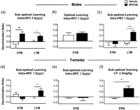
PCAF activation in the HPC enhances STM in males, but not females. a, Under suboptimal learning conditions, where control rats do not remember, PCAF activation (SPV106) within the HPC enhanced STM and LTM compared with vehicle (Veh), in male rats. b, Under optimal learning conditions, PCAF activation did not enhance LTM, in males. c, Under suboptimal learning conditions, intra-PRh PCAF activation enhanced LTM but not STM, in males. d, e, Within females, two high doses of SPV106 within the HPC enhanced LTM but not STM. f, Nevertheless, intraperitoneal (I.P.) administration of SPV106 did enhance STM in females. Data are mean discrimination ratio ± SEM (*p ≤ 0.05; **p ≤ 0.01; corrected for multiple comparisons).
Successful OiP memory additionally relies on PRh in order to process object identity (Winters et al., 2004; Barker and Warburton, 2011; Mitchnick et al., 2015, 2016). We previously reported that PCAF was necessary in male PRh for LTM, but not STM (Mitchnick et al., 2016). Accordingly, activating PCAF in male PRh with SPV106 enhanced LTM, but not STM (Fig. 3c). Specifically, the interaction term (F(1,10) = 1.41; p = 0.262; n = 11) and main effect of delay (F(1,10) = 1.71; p = 0.221) were both nonsignificant, whereas the main effect of drug condition (F(1,10) = 5.29; p = 0.44) was significant. Planned comparisons demonstrated that memory in the SPV106 condition was significantly greater than the vehicle condition for the LTM (t(10) = 7.53; p < 0.001) retention delay, but not the STM (t(10) = 0.46; p = 0.661) retention delay. These results indicate the selective role of PCAF in STM for the dHPC-dependent component of the OiP task in male rats. Given the lack of involvement of PCAF in PRh-mediated OiP memory, the rest of the experiments focused exclusively on the HPC.
In females, while PCAF activation enhanced LTM, no effects were found for STM (Fig. 3d); specifically, the delay × condition interaction (F(1,9) = 0.23; p = 0.643; n = 10) and main effect of retention delay (F(1,9) = 2.34; p = 0.161) were both nonsignificant, whereas the main effect of drug condition was significant (F(1,9) = 16.74; p = 0.003). A significant difference was seen between drug conditions under a LTM retention delay (t(9) = 2.85; p = 0.030). While significant differences were also apparent between drug conditions under a STM retention delay (t(9) = 2.58; p = 0.030), neither condition was significantly different from chance (vehicle, t(9) = 1.37; p = 0.270; SPV106, t(9) = −1.75; p = 0.228), suggesting that PCAF activation within the HPC does not enhance STM in females. Similar results were seen with a higher dose (1.5 μg/μl) of SPV106 (Fig. 3e; paired t test, t(9) = 0.29; p = 0.776). Despite the lack of intra-HPC effects, systemic administration of SPV106 (2.5 mg/kg) did enhance STM in females (Fig. 3f; t(9) = 4.09; p = 0.003), similar to findings in young female mice (Creighton et al., 2022). As such, the remaining experiments focused on characterizing the unique involvement of PCAF in the male HPC.
Intrahippocampal ERα/β inhibition impairs STM and LTM in males
Prior to investigating a functional interaction between PCAF and ERs, we first established whether ERs in the male dHPC are necessary for short-term OiP memory. The OiP task (Fig. 1) and intra-dHPC infusion parameters (Table 1) used were similar to our previous work (Mitchnick et al., 2016). Under normal learning conditions, a high dose (0.5 μg/μl) of the ERα/β inhibitor ICI-182-780 impaired both STM and LTM; specifically, the delay × condition interaction was nonsignificant (F(1,10) = 0.47; p = 0.509; n = 11), whereas both the main effect of delay (F(1,10) = 5.80; p = 0.037) and condition (F(1,10) = 34.17; p < 0.001) were significant. Moreover, the drug condition was significantly impaired compared with the control condition within the STM (t(10) = 4.96; p = 0.002) and LTM (t(10) = 2.88; p = 0.016) retention delays (Fig. 4a), indicating that ERs are necessary in the male rat dHPC for OiP memory. Intra-HPC administration of a low dose (0.1 μg/μl) of the ERα/β inhibitor ICI-182-780 did not impair short-term OiP memory when compared with vehicle (t(10) = 0.29; p = 0.781; Fig. 4b), providing a subeffective dose to use in subsequent interaction experiments.

dHPC ERα/β inhibition impairs STM and LTM in males. a, Under optimal (normal) learning conditions, inhibition of ERα/β in the dHPC by ICI-182-780 impaired STM and LTM. b, A low dose of ICI-182-780 within the dHPC did not impair STM, representing a behaviorally ineffective dose. Data are mean discrimination ratio ± SEM (*p ≤ 0.05; **p ≤ 0.01, corrected for multiple comparisons).
PCAF interacts with ERs in the dHPC to enhance STM, but not LTM, in males
If PCAF and ERs do interact in the dHPC to facilitate memory, inhibiting ERs should block the memory enhancement induced by PCAF activation. A low dose of ICI-182-780 that did not impair memory on its own (Fig. 4b) blocked the STM-enhancing effects of SPV106 under conditions of suboptimal learning (Fig. 5a). Specifically, the significant main effect of drug (F(3,33) = 10.10; p < 0.001; n = 12) was mediated by the enhancing effect of SPV106 compared with vehicle (t(11) = 3.71; p = 0.0045). This effect was blocked by coadministration of a low dose of ICI-182-780 (t(11) = 5.66; p = 0.0015). ICI-182-780 did not affect memory compared with vehicle (t(11) = 1.04; p = 0.319). These results suggest a functional interaction between PCAF and ERs in the dHPC, which facilitates STM.

PCAF and ERs functionally interact to support STM in males. a, b, Under suboptimal learning conditions, a behaviorally ineffective dose of the ERα/β antagonist ICI-182-187 blocked the enhancing effects of PCAF activation on STM, but not LTM, in the dHPC. Veh, vehicle. Data are mean discrimination ratio ± SEM (*p ≤ 0.05; **p ≤ 0.01, corrected for multiple comparisons).
When assessing LTM, the low dose of ICI-182-780 did not affect the memory-enhancing properties of SPV106 under suboptimal learning conditions (Fig. 5b); specifically, the significant main effect of drug (F(3,21) = 12.90; p < 0.001; n = 8) was mediated by the enhancing effects of SPV106 compared with vehicle (t(7) = 3.23; p = 0.042). This effect was not blocked by a low dose of ICI-182-780 (t(7) = 0.09; p = 0.930), and likewise, low-dose ICI-182-780 did not have any effect on memory compared with vehicle (t(7) = 0.499; p = 0.930).
These data indicate that although PCAF and ERs are both necessary for LTM, their mnemonic effects seem to be independent of each other. This might suggest that PCAF acts more similarly to a typical KAT during the consolidation of LTM, modulating gene expression, but potentially engages in a nongenomic ER-mediated signaling pathway to facilitate STM. We chose to focus on this latter, hitherto unreported effect; therefore, the remaining experiments assess only STM.
PCAF interacts specifically with ERα in the dHPC to enhance STM in males
Because ICI-182-780 is an inhibitor of both ERα and ERβ, we are unable to determine from the previous experiment which specific ER(s) PCAF interacts with in the dHPC. Given the paucity of truly selective ERα and ERβ inhibitors, we employed the selective ERα and ERβ activators, PPT (propylpyrazoletriol) and DPN (diarylpropionitrile), respectively. Under suboptimal learning conditions, neither compound at high doses facilitated LTM; memory in the PPT condition did not differ from the vehicle condition (t(9) = 0.58; p = 0.579; n = 10; Fig. 6a), and while memory in the DPN condition was significantly different from the vehicle condition (t(9) = 2.40; p = 0.040; n = 10), neither group demonstrated memory above chance (t(9) = −1.13; p = 0.287 and t(9) = 0.11; p = 0.898, for the control and DPN conditions, respectively; Fig. 6b). With respect to STM, under suboptimal learning conditions a high dose of PPT enhanced STM compared with vehicle (t(9) = 2.82; p = 0.020; n = 10; Fig. 6c), but a high dose of DPN did not (t(9) = 1.23; p = 0.250; Fig. 6d).

PCAF and ERα functionally interact to enhance STM in males. a, b, Under suboptimal learning conditions, neither the ERα agonist PPT nor the ERβ agonist DPN enhanced LTM. c, d, Under suboptimal learning conditions, PPT but not DPN enhanced STM. e, Under optimal learning conditions, a low dose of the PCAF inhibitor embelin did not impair STM, representing a behaviorally ineffective dose. f, Under suboptimal learning conditions, the behaviorally ineffective dose of embelin blocked the STM-enhancing effects of ERα agonism by PPT. Veh, vehicle. Data are mean discrimination ratio ± SEM (**p ≤ 0.01; corrected for multiple comparisons).
If PCAF functionally interacts with ERα, inhibiting PCAF should block the memory-enhancing effects of ERα activation. We used the selective PCAF inhibitor embelin (Table 1), which we previously have shown to impair dHPC-dependent short–term and long–term OiP memory (Mitchnick et al., 2016). Under suboptimal learning conditions, embelin blocked the STM-enhancing effects of PPT (Fig. 6f); specifically, the significant main effect of condition (F(3,27) = 7.27; p = 0.001; n = 10) was mediated by the PPT-induced memory enhancement, in comparison with the vehicle condition (t(9) = 6.17; p = 0.003). This effect was blocked by a low dose (0.1 μg/μl) of embelin (t(9) = 4.59; p = 0.003) that did not affect STM memory on its own under optimal learning conditions (t(9) = 0.03; p = 0.975; Fig. 6e). These results demonstrate that PCAF functionally interacts specifically with ERα in the dHPC to enhance short-term OiP. Moreover, given that these compounds were administered immediately before and after the learning phase and thus were active specifically during encoding/consolidation, it suggests that the STM-enhancing effects of PCAF activation by SPV106 were also due to activity during encoding/consolidation and not due to earlier genomic effects (i.e., given that SPV106 was administered 3 d prior to learning).
The STM-enhancing effects of the PCAF–ERα interaction does not require local estrogen biosynthesis in males
Given that our results suggest that PCAF interacts with ERα within the dHPC, it is worthwhile to determine whether estrogens are also necessary for this functional interaction. To this end, we blocked the aromatization of testosterone into estradiol in the dHPC using the selective aromatase inhibitor letrozole. A dose response experiment demonstrated a main effect of dose (vehicle, 3.0, 4.0, 5.0, and 6.0 μg/μl) on STM under optimal learning conditions (F(3,27) = 4.28; p = 0.014; n = 10). Specifically, when compared with the vehicle condition, memory was significantly worse following 6.0 μg/μl of letrozole (t(9) = 3.69; p = 0.020), but not 3.0 μg/μl (t(9) = 0.816; p = 0.269), 4.0 μg/μl (t(9) = 0.816; p = 0.269), or 5.0 μg/μl (t(9) = 0.816; p = 0.269). Compared with chance, infusions of vehicle (t(9) = 9.29; p < 0.001), 3.0 μg/μl (t(9) = 5.57; p < 0.001), or 4.0 μg/μl (t(9) = 6.52; p < 0.001) of letrozole did not impair memory, while 5.0 μg/μl (t(9) = 0.95; p = 0.459) and 6.0 μg/μl (t(9) = 0.19; p = 0.857; Fig. 7a) doses did, demonstrating that local estrogen biosynthesis is necessary in the male rat dHPC to support short-term OiP memory.

PCAF–ERα interaction does not require local estrogen synthesis to enhance STM in males. a, Under optimal learning conditions, higher doses (5.0 and 6.0 μg/μl) of the aromatase inhibitor letrozole impaired STM compared with chance, while lower doses (3.0 and 4.0 μg/μl) did not. b, Employing the highest behaviorally ineffective dose of letrozole (4.0 μg/μl), the STM-enhancing effects of PCAF activation was not blocked by letrozole. Veh, vehicle. Data are mean discrimination ratio ± SEM (*p ≤ 0.05; ***p < 0.001; corrected for multiple comparisons).
Following this, we investigated the effects of estrogen biosynthesis inhibition on the enhancing effects of PCAF activation. Under suboptimal learning conditions, the highest behavioral ineffective dose of letrozole (4.0 μg/μl) did not block the STM-enhancing effects of PCAF activation by SPV106 (Fig. 7b); specifically, the significant main effect of condition (F(3,27) = 9.94; p < 0.001; n = 10) was mediated by the SPV106-induced memory enhancement, in comparison with the vehicle condition (t(9) = 3.65; p = 0.016). This memory enhancement was not blocked by letrozole in comparison with the vehicle condition (t(9) = 0.46; p = 0.654). Similarly, letrozole did not have any effect on memory, in comparison with the vehicle condition (t(9) = 1.00; p = 0.515). These findings suggest that locally produced estrogens are not necessary for the memory-enhancing properties of this PCAF–ERα interaction in the dHPC.
PCAF activation leads to rapid postlearning phosphorylation of JNK and ERK in males
Enhancing the activity of ERs has been shown to activate various downstream cell signaling pathways involved in LTM and STM, including phosphorylated ERK and JNK (Frick et al., 2018; Sheppard et al., 2023). If PCAF activates ERα to rapidly enhance OiP memory, enhancements of PCAF could cause increases in one or more signaling kinases, postlearning. Therefore, we assessed P-ERK and P-JNK levels in the dHPC of rats that received intra-dHPC PCAF activation (SPV106; high dose) or vehicle, followed by a suboptimal short-term OiP sample phase (2 min; Fig. 8a). In an additional group of rats, coadministration of the PCAF activator (SPV106; high dose) and ERα/ERß inhibitor (ICI-182-780; behaviorally ineffective low dose) provided an opportunity to block any potential enhancements in P-ERK and/or P-JNK.

PCAF activation enhances estrogen signaling-related kinase activation in males. a, Experimental flow for each group. b, Representative blots for P-JNK p54 p46 (top; activated) and total JNK p54 and p46 (bottom). c, Intra-HPC PCAF activation (intra-HPC SPV106 infusion) enhanced P-JNK (collapsed across isoforms) following a suboptimal OiP sample phase (2 min), compared with a vehicle infusion. A behaviorally ineffective (low) dose of the ERα/β antagonist ICI-182-780 did not abolish the SPV-106 increases in P-JNK. d, Representative blots for P-ERK1/2 (top; activated) and total ERK1/2 (bottom). e, Intra-HPC PCAF activation (intra-HPC SPV106 infusion) elevated P-ERK1/2 (collapsed across isoform) following a suboptimal OiP sample phase (2 min), although this effect did not reach significance. A behaviorally ineffective dose of the ERα/β antagonist ICI-182-780 did not abolish the SPV106 increases in P-JNK. Veh-Veh, vehicle + vehicle group; SPV-Veh, SPV106 + vehicle group; SPV-ICI, SPV106 + ICI-182-780 group. Data represent mean ± SEM phosphorylated/total protein relative to the veh-veh group (*p ≤ 0.05, corrected for multiple comparisons).
Five minutes following object exposure, P-JNK levels in dHPC were increased in the context of PCAF activation. Specifically, a significant effect of treatment on JNK phosphorylation was detected using a two-way ANOVA (F(2,36) = 3.32; p = 0.047; n = 21), with no significant differences in JNK isoform (F(1,36) = 0.47; p = 0.498) or interaction between treatment and JNK isoform (F(1,36) = 0.47; p = 0.498). Planned comparisons, collapsing across isoforms (i.e., ERK1/2; JNK 54kDA/46kDA), indicated that intra-dHPC SPV106 significantly increased JNK phosphorylation compared with vehicle infusion (t(26) = 2.67; p = 0.026). This effect was not abolished by ICI-182-780 (low dose), remaining significantly increased compared with the vehicle group (t(26) = 2.44; p = 0.022; Fig. 8b,c).
In terms of P-ERK levels, the interaction was not significant (F(2,36) = 0.21; p = 0.812; n = 21), and there was no effect of isoform (F(1,36) = 0.30; p = 0.589). The effect of treatment on ERK phosphorylation was also nonsignificant (F(2,36) = 2.77; p = 0.076), despite a noticeable increase in the SPV106 condition (Fig. 6d,e). Planned comparisons, collapsing across isoform, indicated that intra-dHPC SPV106 increased ERK phosphorylation compared with vehicle infusion (t(26) = 2.35; p = 0.053), although not significantly so. Coadministration of SPV106 and ICI-182-780 (low dose) significantly increased ERK phosphorylation compared with the vehicle group (t(26) = 2.44; p = 0.022; Fig. 6d,e). These results might suggest that PCAF activation is leading to increases in P-ERK, as it is less likely that ERα/ERß inhibition would increase related downstream signaling kinases.
Discussion
The present findings reveal a novel functional, and potentially estrogen-independent, interaction between PCAF and ERα in the dHPC of male rats. The dual demonstration of this interaction (i.e., blocking the STM-enhancing effects of PCAF activation with ER inhibition and blocking the STM-enhancing effects of ERα activation with selective PCAF inhibition) strengthens this interpretation. Given that the compounds used in one of these interaction experiments were administered/active at the time of learning suggests that the PCAF–ERα interaction is occurring rapidly at the nongenomic level, to facilitate STM. Moreover, STM is not impaired when PCAF is perturbed at the genomic level (i.e., via siRNAs; Mitchnick et al., 2016). Our results further suggest that this PCAF–ERα interaction may be specific to dHPC-dependent STM of males, as it was not found at longer delays and was not seen within the PRh, and no such interaction was present between PCAF and ERβ. Additionally, PCAF activation increased P-JNK and P-ERK, downstream ER signaling kinases, beyond levels induced by suboptimal OiP training, suggesting a possible mechanism by which PCAF exerts rapid beneficial effects on HPC-dependent STM. Finally, this HPC-dependent mechanism appears to be unique to males, as it was not found in females.
PCAF acts as an epigenetic and nonepigenetic regulator
The long-term OiP memory enhancement by PCAF activation (via SPV106) in the dHPC of males and females (and in the PRh) is consistent with studies demonstrating the necessity of PCAF (Maurice et al., 2008; Mitchnick et al., 2016) and the promnemonic effects of PCAF activation (Wei et al., 2012) on LTM. In addition to enhancing PCAF, SPV106 simultaneously inhibits CBP (CREB-binding protein), another HAT (Sbardella et al., 2008). As such, the finding that SPV106 can lead to LTM enhancements is interesting in light of a body of literature suggesting the necessity of CBP in the HPC for the beneficial effects of HDAC inhibition. In opposition to HATs, HDACs catalyze the removal of acetyl groups and the condensation of chromatin, inducing a state of gene repression. Many studies have shown that HDAC inhibition has promnemonic effects by facilitating gene expression (Guan et al., 2009; Stefanko et al., 2009; Kilgore et al., 2010; McQuown et al., 2011; Rogge et al., 2013; Valiati et al., 2017; Shu et al., 2018), but for HPC-dependent object recognition, this effect has been shown to require functional CBP (Vecsey et al., 2007; Roozendaal et al., 2010; Haettig et al., 2011; McQuown et al., 2011). Moreover, our lab and others have demonstrated CBP inhibition, mRNA reduction, or knock-out to impair LTM on a variety of tasks (Oike et al., 1999; Alarcón et al., 2004; Wood et al., 2005, 2006; Barrett et al., 2011; Haettig et al., 2011; Valor et al., 2011; Mitchnick et al., 2016). Therefore, finding that PCAF activation by SPV106 is capable of enhancing HPC-dependent memory despite its inhibition of CBP suggests the compound's potential use therapeutically, such as in Rubinstein–Taybi syndrome, which is characterized by craniofacial abnormalities and moderate-to-severe intellectual disabilities due to CBP mutations (Oike et al., 1999). In contrast, pharmacological inhibition or mRNA reduction of CBP does not affect STM (Korzus et al., 2004; Barrett et al., 2011; Chatterjee et al., 2013; Maddox et al., 2013a,b; Stilling et al., 2014; Mitchnick et al., 2016). Furthermore, the PCAF inhibitor (embelin) used in the present study does affect CBP. Collectively, these suggest that the enhancing effects of SPV106 on STM in the current study were due entirely to its activation of PCAF, not its inhibition of CBP, although an agent that only enhances PCAF would show this definitively (Castellano et al., 2015).
With respect to the PCAF–ERα interplay demonstrated in the current study, other groups have also demonstrated interactions between PCAF and nonhistone proteins in the regulation of memory, albeit LTM (Wei et al., 2012; Park et al., 2015). As discussed, epigenetic mechanisms are typically only implicated in LTM, as they regulate gene expression that is not necessary for STM (Sheppard et al., 2021, 2023). For example, pharmacological inhibition or activation of KATs, such as CBP or p300, during learning consistently cause LTM (24 h) but not STM (<4 h) impairments or enhancements, respectively (Korzus et al., 2004; Barrett et al., 2011; Chatterjee et al., 2013; Maddox et al., 2013a,b; Stilling et al., 2014; Mitchnick et al., 2016). While STM impairments have been seen in KO mouse models of PCAF (Maurice et al., 2008), CBP (G. Chen et al., 2010), and HDAC2 (Guan et al., 2009), as well as in flies with developmental genetic reduction of HDAC6 or the KAT Tip60 (Xu et al., 2014; Perry et al., 2017), these models make it more difficult to draw conclusions in light of possible developmental compensatory changes and the inability to specifically target the learning/consolidation phase of memory. Nevertheless, inhibition of PCAF specifically during learning (i.e., via pharmacological inhibition) impairs short-term (5 min, 20 min, 3 h) object memory in rats and mice (Mitchnick et al., 2016; Creighton et al., 2022). Here we also demonstrate that PCAF is necessary for the STM enhancements caused by immediate postlearning ERα agonism. Collectively, these latter findings suggest that PCAF is most likely acting through a nonepigenetic mechanism to facilitate STM (but this remains to be demonstrated directly).
ERs are involved in HPC-dependent OiP memory in males
The impairing effect of ERα/ERβ antagonism on OiP memory is consistent with the known role of ERα/ERβ in HPC-dependent memory (Sheppard et al., 2018). The STM impairments were likely due to antagonism of ERα specifically, given that intra-dHPC ERα, but not ERβ, agonism facilitated STM. Indeed, previous studies have demonstrated enhancements in short-term object memories following intra-dHPC ERα agonism in both female ovariectomized mice (Phan et al., 2015) and rats (Walf et al., 2006; Frye et al., 2007) and long-term object location memory impairments in female ovariectomized mice following intra-dHPC ERα antagonism (Kim and Frick, 2017). Here we show an involvement of ERα in dHPC-dependent memory in males, as well.
Interestingly, activating ERβ, but not ERα, in PRh of intact male rats enhanced long-term OiP memory, while no effect of intra-PRh ERα/ERβ inhibition was seen on STM (Mitchnick et al., 2019). This demonstrates regional differences in ER involvement in males for different forms of memory, similar to previous reports (Phan et al., 2011, 2015; Kim and Frick, 2017), further emphasizing the complexity of estrogen/ER effects on memory.
PCAF functionally interacts with ERα in males to enhance STM
A few studies have indicated the intersection of epigenetic mechanisms and estrogen signaling for memory formation. Specifically, intra-dHPC administration of 17β-estradiol coupled with object learning increased acetylation at specific histone sites, including around the BDNF promoter, in addition to decreasing HDAC2 and HDAC3 protein levels (Z. Zhao et al., 2010, 2012; Fortress et al., 2014). Furthermore, some of these effects were blocked by p300 HAT inhibition, as was long-term object memory (Z. Zhao et al., 2012). In the current study, PCAF activation presumably acetylated histone proteins to enhance gene expression and LTM. Nevertheless, the PCAF–ERα interaction supporting STM presented here indicates dissociable roles of PCAF in HPC-mediated LTM and STM. PCAF has previously been shown to activate ERα and ERRα in cell culture (Blanco et al., 1998; Wilson et al., 2010), and coinhibition of PCAF and p300 (another lysine acetyltransferase) within the dHPC was shown to abolish the increases in dHPC acetylation of histone 3 by 17β-estradiol (Z. Zhao et al., 2012), but to our knowledge, this is the first demonstration that PCAF and ERα interact in the brain to affect behavior. Furthermore, this illustrates a novel mode of activation of ERα in the rapid facilitation of memory.
Local estrogen synthesis appears dispensable for the STM-enhancing effects of PCAF–ERα in males
Although aromatase inhibition has been shown to cause memory enhancements in a few cases (Cherrier et al., 2005; Moradpour et al., 2006; Alejandre-Gomez et al., 2007), the majority of studies demonstrate impairments in HPC-dependent memory, including in human females (Bayer et al., 2015), ovariectomized female mice (Tuscher et al., 2016), gonadally intact male songbirds (Bailey et al., 2013), gonadally intact mice (J. Zhao et al., 2018; but see Koss and Frick, 2019) and rats (Graham and Milad, 2014), as well as gonadectomized male mice (Koss and Frick, 2019). Moreover, aromatase inhibition impaired short-term and long-term OiP memory in gonadally intact male rats when infused into PRh (Mitchnick et al., 2019). Therefore, the current results agree with many previous findings and indicate that estrogen synthesis is necessary in the gonadally intact male rat dHPC for short-term OiP memory.
Nevertheless, low-dose aromatase inhibition did not affect the STM-enhancing properties of PCAF activation in the dHPC of males, suggesting that PCAF rapidly interacts with ERα in an estrogen-independent manner, although possible effects of circulating hormones from the gonads or other tissues cannot be ruled out (Cui et al., 2013). Interestingly, females with ductal hyperplasia, a premalignant marker for breast cancer, have a mutation in the K303 acetylation site that results in decreased acetylation and increased estrogen sensitivity (Fuqua et al., 2000). The inverse correlation between acetylation on ERα and ligand sensitivity indicates that ERα activation via acetylation can occur in an estrogen-independent manner, as our results might suggest. Moreover, studies have demonstrated the role of ERα in enhanced cognitive function independent of ovarian or exogenously administered estrogens, including via brain-derived neuroestrogens and insulin-like growth factor-1 signaling (Baumgartner and Daniel, 2021). The current results suggest that PCAF-related mechanisms should also be added to this list.
PCAF-induced rapid activation of ER-related signaling kinases following suboptimal learning
Estradiol/ER activity increases the activation of downstream signaling cascades such as ERK (Fernandez et al., 2008; Z. Zhao et al., 2010; Boulware et al., 2013; Fortress et al., 2013; Pereira et al., 2014; Kumar et al., 2015) and JNK (Kim et al., 2016) required for HPC-dependent memory. Moreover, we previously demonstrated that OiP learning rapidly (5 min) increased P-JNK and P-ERK2 in PRh of gonadally intact male rats, in comparison with a nonlearning control group (Mitchnick et al., 2019). In the present study, PCAF activation enhanced P-JNK with a trend toward an increase in P-ERK beyond levels induced by suboptimal OiP training, highlighting signaling mechanisms likely involved in the rapid facilitation of OiP memory in the dHPC of males. Maurice et al. (2008) also noted increased P-ERK in the HPC of both male and female mice immediately following LTM retrieval in a passive avoidance task, but this was not seen in their PCAF knock-out mice (Maurice et al., 2008). Together, these results indicate a clear relationship between PCAF activation and ERK signaling in the HPC of male rats. This finding is particularly interesting in light of a recent report that dHPC administration of estrogens did not cause rapid dHPC enhancement of P-ERK in male mice (Koss et al., 2018), as has been demonstrated in female mice (Fortress et al., 2013; Kim et al., 2016). In light of the current results, this suggests that PCAF might act in the absence of or instead of estrogens to stimulate ERs and subsequently activate ERK, specifically in males.
Despite the functional interaction between PCAF and ERα that was necessary for rapid enhancement of short-term OiP memory in the dHPC of male rats, PCAF-induced elevations in P-JNK and P-ERK were not blocked by ICI-182-780. It is not entirely clear why this is the case. It is possible that, while PCAF could activate many intracellular signaling cascades, it only activates certain kinases upon its interactions with ERα, and we did not assess these specific kinases. Although other ERα/ERβ-related signaling kinases have been identified, many are part of the ERK signaling pathway and might all be expected to show similar activation effects (Frick et al., 2018). Alternatively, there is also the possibility of more complex interactions between ERα and GPER, with some research suggesting that the ERs can work synergistically, antagonistically, in sequence, or in other arrangements (Hadjimarkou and Vasudevan, 2018). Specifically, in addition to blocking ERα/ERβ, ICI-182-780 activates GPER (Hadjimarkou and Vasudevan, 2018), and this could partly explain the unexpected effects with this drug on P-JNK and P-ERK levels. PCAF might also interact with both GPER and ERα to regulate STM. Interestingly, GPER activity has been associated with JNK, but not ERK, signaling in the dHPC and PRh (Kim et al., 2016; Mitchnick et al., 2019). Indeed, PCAF has been shown to interact with GPER (C. Chen et al., 2019). Future studies might aim to determine the regulatory capacity of PCAF more broadly for estrogen signaling in the brain, given our demonstration that dHPC PCAF activation can enhance activity of downstream estrogen signaling kinases beyond levels induced by suboptimal learning parameters, which (presumably) enhances STM.
Sex differences
The STM-enhancing effects of intra-HPC PCAF in males was not seen in females. Nevertheless, brain wide PCAF activation (systemic administration) did enhance short-term OiP memory in female rats, as was documented previously in female mice (Creighton et al., 2022). This might suggest that female rats are utilizing different encoding and/or mnemonic strategies during the OiP task that are less HPC-based. For example, in spatial navigation/spatial memory experiments in rodents or humans, males were more likely to use distal environmental cues to navigate, while females tended to use local landmark cues or even an idiothetic strategy (Bertholet et al., 2014; Yagi et al., 2016; Tronson and Keiser, 2019). Moreover, female rodents have been demonstrated to outperform males in tasks of object recognition (Saucier et al., 2008; Bettis and Jacobs, 2012), with some indication that females are better at discriminating between similar objects (Bettis and Jacobs, 2012; but see alternative results in humans, Aldi et al., 2018). It could be the case that STM enhancement by systemic PCAF activation in female rats is mediated by PCAF's effects within the PRh, possibly exerting influence on ERβ or GPER, given that ERα is sparse or nonexistent in PRh of females (Merchenthaler et al., 2004). Alternatively, the PCAF–ER functional interaction might be a male-specific mechanism of STM enhancement in the context of lower levels of circulating estrogens, particularly given the current findings that PCAF's influence on STM in males appears to be independent of local estrogen synthesis.
Conclusion
The functional interaction between PCAF and ERα that facilitates short-term dHPC–dependent OiP memory suggests a novel substrate for PCAF's promnemonic effects and a novel mechanism for rapid activation of ERs and ER-related signaling kinases that appears to be independent of local estrogen synthesis. Furthermore, this HPC effect was found to be sex-specific, possibly suggesting a male-tailored mechanism to enhance STM. Understanding how PCAF differentially enhances STM in females is a necessary next step. Future work should also look to characterize the nature of a physical interaction between PCAF and ERα, including possible cell-type specificity, given that PCAF (Demyanenko et al., 2020) and ERs (Mhyre and Dorsa, 2006) are both found in neurons and glia. Taken together, the male-specific involvement of PCAF in short-term and long-term HPC–dependent memory and its novel intersection with ERα in the absence of local estrogen synthesis adds to the cognitive effects of (typical) ligand-independent activation of ERα (Baumgartner and Daniel, 2021) and further suggests that PCAF has a much higher therapeutic potential than previously recognized.
Footnotes
- Received August 20, 2023.
- Revision received July 5, 2024.
- Accepted July 15, 2024.
-
This research was supported by National Science and Engineering Research Council (NSERC) of Canada research grants to B.D.W. (400176), N.J.M. (2015-04537), and E.C. (401375). G.S. is supported by grants from the Italian Ministero dell’Istruzione, dell’Università e della Ricerca (MIUR), Progetti di Ricerca di Interesse Nazionale (PRIN 2020CW39SJ), University of Salerno (FARB grant), and Regione Campania (Italy) Grant (Project No. B61G18000470007). Graduate students were supported by NSERC scholarships awarded to K.A.M., C.W., and S.D.C., and an Ontario Graduate Scholarship was awarded to K.N. We thank Paul Sheppard for stimulating discussions and Keon Coleman for brain tissue sectioning and staining.
-
The authors declare no competing financial interests.
- Correspondence should be addressed to Krista A. Mitchnick at kmitchni{at}uoguelph.ca or Boyer D. Winters at bwinters{at}uoguelph.ca.
- Copyright © 2024 the authors




