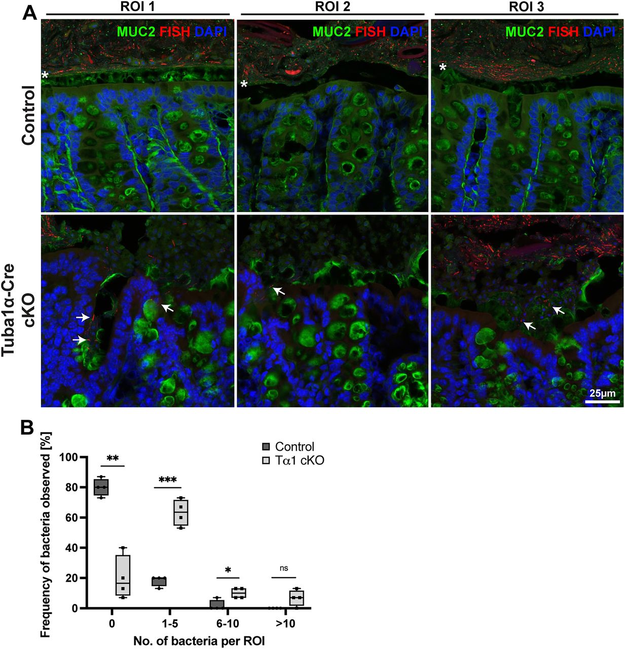
Epithelial barrier disorganization in the adult Tuba1a-Cre;Elp1loxp/loxp cKO mice. A, Representative images of the firm layer of mucin (MUC2) in the gut lumen. MUC2 (green), FISH (red), and DAPI (blue) stains demarcate the mucus layer and bacterial localizations at the border between the lumen and epithelium; n = 3 different control regions of interest (ROI) and Elp1 cKO mice. Asterisks (*) indicate the localization of the firm mucus layer in the gut lumen of the control which is much less obvious in the cKO. Scale bar, 100 µm. B, Bacteria were frequently observed in cKO colon epithelium, and in larger numbers, compared with controls [no bacteria observed, p = 0.002; 1–5 bacteria observed in region of interest (ROI), p = 0.0006; >10 bacteria observed in FOV, p = 0.09]. n = 4 FD mice and 4 controls. White arrows indicate FISH positive bacteria in the mucosa. Scale bars, 100 µm. Error bars, ±SEM. *p ≤ 0.05, **p ≤ 0.01, ***p ≤ 0.001. Each data point represents an individual mouse.


